. 2012 Jan 27;36(1):32-42.
doi: 10.1016/j.immuni.2011.11.015.

Syk kinase-coupled C-type lectin receptors engage protein kinase C-δ to elicit Card9 adaptor-mediated innate immunity

Affiliations

Syk kinase-coupled C-type lectin receptors engage protein kinase C-δ to elicit Card9 adaptor-mediated innate immunity

Dominikus Strasser et al. Immunity. .

Abstract

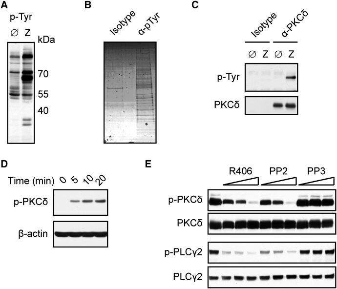
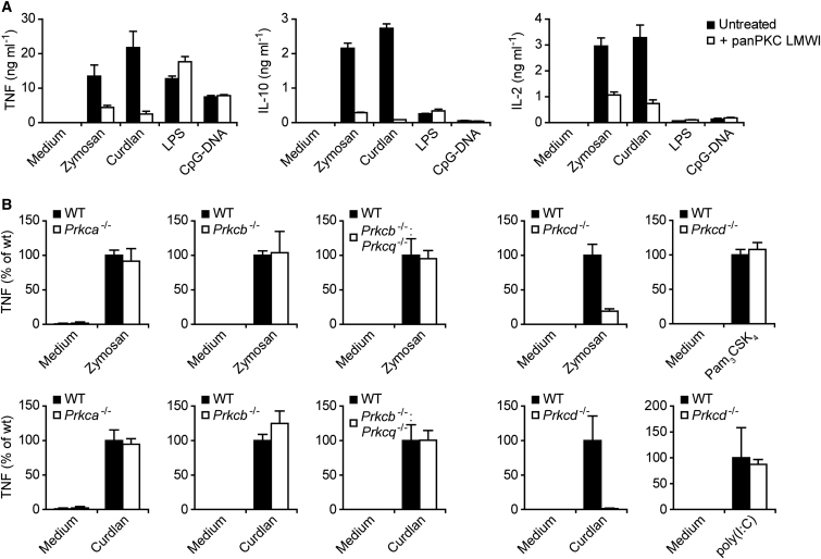
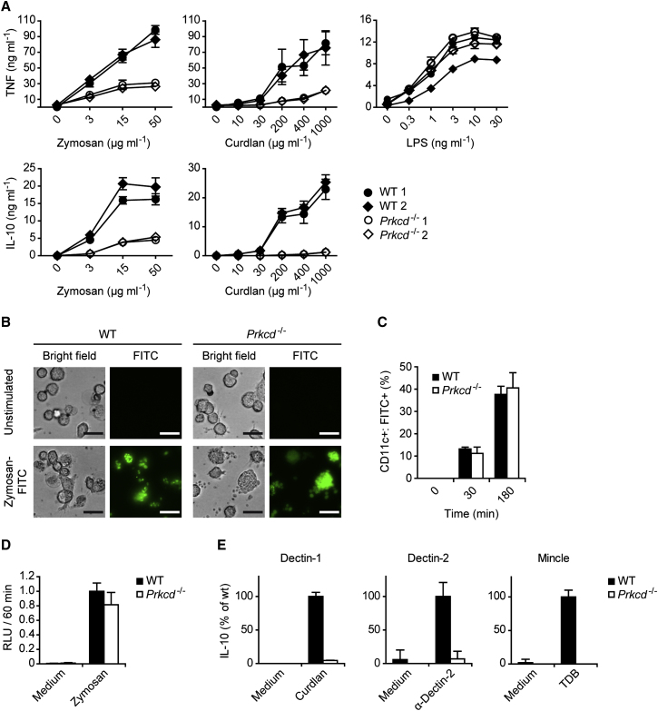
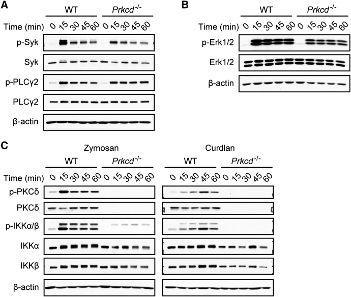
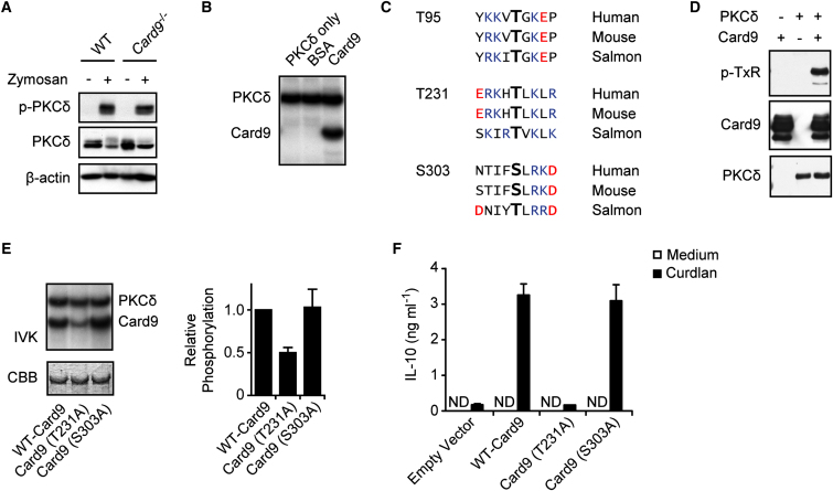
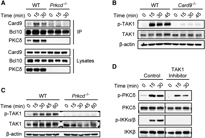
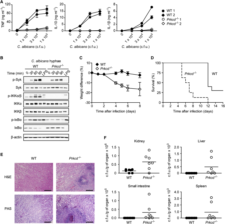
C-type lectin receptors (CLRs) that couple with the kinase Syk are major pattern recognition receptors for the activation of innate immunity and host defense. CLRs recognize fungi and other forms of microbial or sterile danger, and they induce inflammatory responses through the adaptor protein Card9. The mechanisms relaying CLR proximal signals to the core Card9 module are unknown. Here we demonstrated that protein kinase C-δ (PKCδ) was activated upon Dectin-1-Syk signaling, mediated phosphorylation of Card9 at Thr231, and was responsible for Card9-Bcl10 complex assembly and canonical NF-κB control. Prkcd(-/-) dendritic cells, but not those lacking PKCα, PKCβ, or PKCθ, were defective in innate responses to Dectin-1, Dectin-2, or Mincle stimulation. Moreover, Candida albicans-induced cytokine production was blocked in Prkcd(-/-) cells, and Prkcd(-/-) mice were highly susceptible to fungal infection. Thus, PKCδ is an essential link between Syk activation and Card9 signaling for CLR-mediated innate immunity and host protection.


Figures

Figure 1
Figure 1
PKCδ Is Tyrosine Phosphorylated upon Dectin-1 Ligation (A) BMDCs were left untreated (Ø) or stimulated for 10 min with zymosan (Z). Cellular lysates were immunoblotted with phospho-Tyrosine (p-Tyr) antibodies. (B) BMDCs were stimulated with zymosan as in (A). Proteins were immunopurified with p-Tyr antibodies or IgG1 control (isotype) and visualized by Coomassie staining after electrophoresis. (C) BMDCs were left untreated or stimulated with zymosan as in (A). Proteins from lysates were immunoprecipitated with PKCδ antibodies or isotype control and analyzed by immunoblot with p-Tyr or PKCδ antibodies. (D) Immunoblot analysis of BMDCs stimulated with zymosan for various times and probed with antibodies against phospho-PKCδ (Tyr311) or β-actin. (E) BMDCs were left untreated or preincubated with Syk inhibitor R406 (0.5, 1, 2 μM), Src-kinase inhibitor PP2 (1.5, 3, 6 μM), or its inactive analog PP3 (1.5, 3, 6 μM) and stimulated for 10 min with zymosan. Lysates were analyzed by immunoblot with antibodies against phospho-PKCδ, PKCδ, phospho-PLCγ2, or PLCγ2. All results are representative of at least three independent experiments.
Figure 2
Figure 2
Selectively Impaired Dectin-1 Signaling in Prkcd−/− BMDCs (A) BMDCs were left untreated or preincubated with PKC inhibitor panPKC LMWI (5 μM) and stimulated with zymosan (20 μg ml−1), curdlan (400 μg ml−1), LPS (200 ng ml−1), or CpG-DNA (2 μM) for 6 hr. TNF, IL-10, and IL-2 concentrations in the supernatants were assayed by ELISA. Data are expressed as means + SD of triplicate samples. (B) TNF production in Prkca−/−, Prkcb−/−, Prkcq−/−, or Prkcd−/− BMDCs that were left untreated (Medium) or stimulated with zymosan (20 μg ml−1), curdlan (200 μg ml−1), Pam3CSK4 (30 ng ml−1), or poly(I:C) (30 μg ml−1) for 6 hr as indicated. Data are expressed as percent of WT + SD, derived from stimulations in triplicates.
Figure 3
Figure 3
PKCδ Deficiency Particularly Impairs CLR Signaling but Not Phagocytosis (A) WT and Prkcd−/− BMDCs were incubated with the indicated concentrations of zymosan, curdlan, or LPS. TNF and IL-10 concentrations in the supernatants were assayed by ELISA. Data are expressed as means ± SD of triplicates. (B) BMDCs from WT and Prkcd−/− mice were incubated for 2 hr with FITC-zymosan particles (100 μg ml−1). FITC-zymosan internalization was visualized by fluorescence microscopy (scale bars represent 20 μm). (C) BMDCs were incubated with FITC-zymosan as in (B) for the times indicated. The frequencies of CD11c+ cells containing zymosan-FITC particles were quantified by FACS analysis. (D) BMDCs from WT and Prkcd−/− mice were incubated with Luminol and were left untreated (Medium) or stimulated with zymosan. Luminescence was assayed for 60 min in 10 min intervals and relative light units (RLU) as a measure for ROS generation integrated over time. Results represent means + SD from three independent experiments. (E) WT and Prkcd−/− BMDCs were stimulated through Dectin-1, Dectin-2, or Mincle with curdlan (20 μg ml−1), plate-bound Dectin-2 antibody, or TDB (100 μg ml−1), respectively. IL-10 concentrations in the cell culture supernatants were quantified by ELISA. Data are expressed as percent of WT + SD, derived from stimulations in triplicates. All results are representative of at least three independent experiments.
Figure 4
Figure 4
PKCδ Controls NF-κB Activation (A) Regular Syk and PLCγ2 activation in Prkcd−/− cells. BMDCs from WT or Prkcd−/− mice were stimulated with zymosan for the indicated times. Syk and PLCγ2 activation was determined by immunoblot with phospho-Syk or phospho-PLCγ2 antibodies. Immunoblotting with Syk, PLCγ2, and β-actin antibodies indicate equal protein loading. (B) WT or Prkcd−/− BMDCs were stimulated with zymosan as indicated. Activation of the MAP kinases Erk1 and Erk2 was determined by immunoblot with phospho-Erk1/2 antibodies. Immunoblotting with Erk1/2 and β-actin antibodies indicates equal protein loading. (C) Defective NF-κB signaling in Prkcd−/− BMDCs. Cells were stimulated with zymosan or curdlan as indicated. Lysates were analyzed by immunoblot with phospho-PKCδ, phospho IKKα/β and PKCδ, IKKα, IKKβ, and β-actin antibodies.
Figure 5
Figure 5
PKCδ Mediates Card9 Phosphorylation for Cytokine Production (A) Normal PKCδ activation in Card9−/− cells. BMDCs from WT and Card9−/− mice were stimulated for 10 min with zymosan. PKCδ activation was determined by immunoblot with antibodies against phospho-PKCδ, PKCδ, or β-actin. (B) In vitro kinase assay. Recombinant PKCδ was incubated with recombinant Card9 or BSA and γ-[32P]ATP for 60 min. Autophosphorylation of PKCδ and phosphorylation of Card9 was visualized by autoradiography. (C) Alignment of highly conserved putative PKC phosphorylation sites within Card9. Basic amino acids are depicted in blue and acidic amino acids in red. (D) Recombinant PKCδ and/or recombinant Card9 was incubated with ATP for 2 hr. Card9 TxR-phosphorylation was determined by immunoblot with antibodies against phospho-TxR, Card9, or PKCδ. (E) Strep-tagged WT-Card9, Card9(T231A), or Card9(S303A) were expressed in HEK293 cells, purified, and incubated in vitro with recombinant PKCδ and γ-[32P]ATP. Left: phosphorylation of PKCδ and Card9 mutants were analyzed by autoradiography. Equal Card9 loading was verified by Coomassie Brilliant Blue (CBB) staining. Right: Card9 phosphorylation quantified by densitometry. Results represent means + SD from three independent experiments. (F) Card9-deficient BMDCs were retrovirally reconstituted with empty vector or vectors expressing WT-Card9, Card9(T231A), or Card9(S303A). Reconstituted cells were stimulated with Curdlan and IL-10 concentrations in the supernatants were measured by ELISA. ND: not detectable. Results represent means + SD from one experiment representative of three independent experiments.
Figure 6
Figure 6
PKCδ Triggers Card9-Dependent TAK1 Activation (A) PKCδ controls Card9-Bcl10 complex assembly. WT and Prkcd−/− BMDCs were stimulated with zymosan for the indicated times and lysates subjected to immunoprecipitation with Bcl10-specific antibodies. Immunoprecipitates and total lysates were immunoblotted as indicated. (B) Card9-dependent TAK1 activation. WT or Card9−/− cells were stimulated with zymosan as indicated. Lysates were immunoblotted with antibodies against phospho-TAK1, TAK1, or β-actin. (C) PKCδ mediates TAK1 activation. BMDCs from WT or Prkcd−/− mice were stimulated as in (B). Lysates were analyzed by immunoblot with antibodies against phospho-TAK1, TAK1, or β-actin. (D) TAK1 signaling is critical for IKK activation. WT BMDCs were pretreated for 30 min with DMSO (control) or the selective TAK1 inhibitor (5Z)-7-Oxozeaenol (2 μM) and stimulated with zymosan. Lysates were analyzed by protein immunoblotting for PKCδ and IKK activation with antibodies against p-PKCδ, PKCδ, p-IKKα/β, and IKKβ.
Figure 7
Figure 7
PKCδ Is Critical for Antifungal Host Defense (A) BMDCs from WT and Prkcd−/− mice were incubated with increasing doses of live C. albicans hyphae. Concentrations of TNF, IL-10, and IL-1β in the culture supernatants were determined 6 hr later. Results are means ± SD of triplicates. (B) BMDCs were stimulated for the indicated times with C. albicans hyphae or for 30 min with 100 ng ml−1 LPS. Lysates were analyzed by immunoblot with antibodies against phospho-Syk, Syk, phospho-IKKα/β, IKKα, IKKβ, phospho-IκBα, IκBα, or β-actin. (C and D) WT (n = 9) and Prkcd−/− (n = 8) mice were infected intravenously with 5 × 104 colony-forming units (c.f.u.) of C. albicans and monitored daily for weight changes (C) and survival (D). Statistical analysis was performed by log-rank test (p < 0.0001). (E) Kidney sections from C. albicans-infected WT and Prkcd−/− mice were stained with H&E or PAS (scale bars represent 300 μm). (F) Mice were infected intravenously with 1 × 104 c.f.u. of C. albicans. After 8 days, C. albicans titers were determined in the kidneys, the liver, the small intestine, and the spleen.

References

    1. Ariizumi K., Shen G.L., Shikano S., Xu S., Ritter R., 3rd, Kumamoto T., Edelbaum D., Morita A., Bergstresser P.R., Takashima A. Identification of a novel, dendritic cell-associated molecule, dectin-1, by subtractive cDNA cloning. J. Biol. Chem. 2000;275:20157–20167. - PubMed
    1. Blom N., Sicheritz-Pontén T., Gupta R., Gammeltoft S., Brunak S. Prediction of post-translational glycosylation and phosphorylation of proteins from the amino acid sequence. Proteomics. 2004;4:1633–1649. - PubMed
    1. Brown G.D., Gordon S. Immune recognition. A new receptor for beta-glucans. Nature. 2001;413:36–37. - PubMed
    1. Chen S.T., Lin Y.L., Huang M.T., Wu M.F., Cheng S.C., Lei H.Y., Lee C.K., Chiou T.W., Wong C.H., Hsieh S.L. CLEC5A is critical for dengue-virus-induced lethal disease. Nature. 2008;453:672–676. - PubMed
    1. Choudhary C., Olsen J.V., Brandts C., Cox J., Reddy P.N., Böhmer F.D., Gerke V., Schmidt-Arras D.E., Berdel W.E., Müller-Tidow C. Mislocalized activation of oncogenic RTKs switches downstream signaling outcomes. Mol. Cell. 2009;36:326–339. - PubMed

Publication types

MeSH terms