. 2012 Mar 29:206:60-8.
doi: 10.1016/j.neuroscience.2011.12.040. Epub 2012 Jan 4.

Development of the sigma-1 receptor in C-terminals of motoneurons and colocalization with the N,N'-dimethyltryptamine forming enzyme, indole-N-methyl transferase

Affiliations

Development of the sigma-1 receptor in C-terminals of motoneurons and colocalization with the N,N'-dimethyltryptamine forming enzyme, indole-N-methyl transferase

T A Mavlyutov et al. Neuroscience. .

Abstract

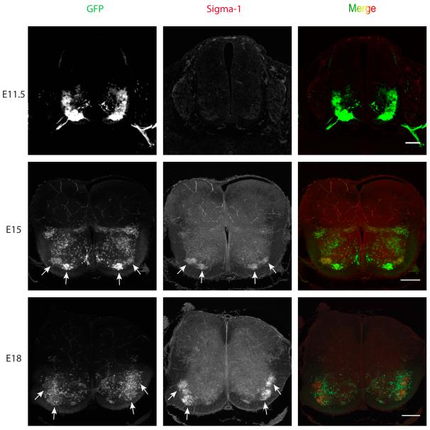
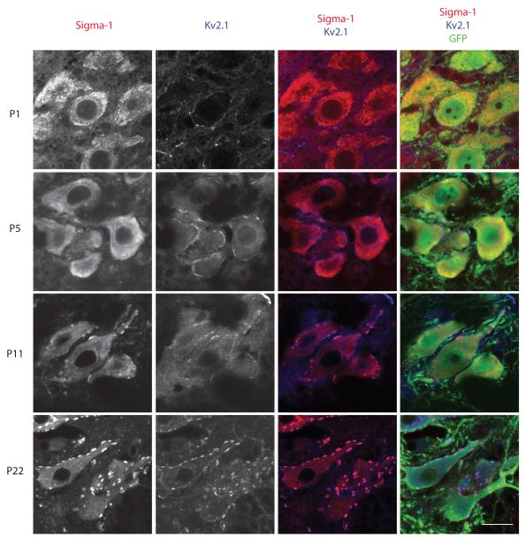
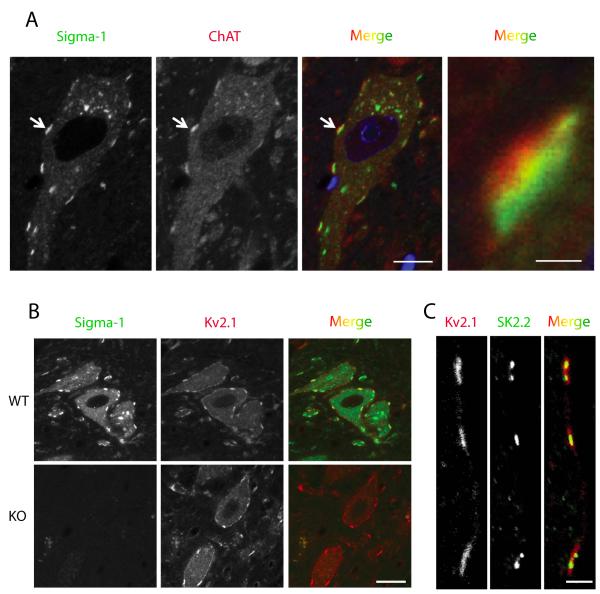
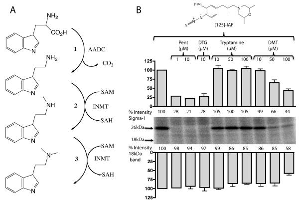
The function of the sigma-1 receptor (S1R) has been linked to modulating the activities of ion channels and G-protein-coupled receptors (GPCR). In the CNS, the S1R is expressed ubiquitously but is enriched in mouse motoneurons (MN), where it is localized to subsurface cisternae of cholinergic postsynaptic densities, also known as C-terminals. We found that S1R is enriched in mouse spinal MN at late stages of embryonic development when it is first visualized in the endoplasmic reticulum. S1Rs appear to concentrate at C-terminals of mouse MN only on the second week of postnatal development. We found that indole-N-methyl transferase (INMT), an enzyme that converts tryptamine into the sigma-1 ligand dimethyltryptamine (DMT), is also localized to postsynaptic sites of C-terminals in close proximity to the S1R. This close association of INMT and S1Rs suggest that DMT is synthesized locally to effectively activate S1R in MN.


Figures

Figure 1
Figure 1
Development of the Sigma-1 receptor in the embryonic mouse spinal cord. Sigma-1 receptor immunostaining is not visible in the mouse MN at embryonic stage E11.5. At E15 the Sigma-1 receptors concentrated in MNs in the lateral and medial motor columns in upper lumbar segments of the spinal cord. Note that not all MNs in the lateral column are GFP positive. The expression of S1Rs increased at E18 in the lateral motor column in the lower lumbar segments of the spinal cord. Not all MNs express GFP. Transgenic HB9-derived GFP mice were used for identification of MNs. Arrows point to clusters of ventral MNs. Scale bar = 100μm.
Figure 2
Figure 2
Development of the Sigma-1 receptor in GFP-expressing MN in the spinal cord of the transgenic Hb9-GFP mouse. Immunohistochemical staining of the Sigma-1 receptor is difuse at early postnatal ages (P1, P5). However, an overlay of the staining of sigma-1 receptors and Kv2.1 that identifies the C-terminals demonstrated that the receptor is concentrated in C-terminals (purple) by postnatal day P11. Scale bar = 20μm.
Figure 3
Figure 3
Adult mouse MN. The Sigma-1 receptor is found in C-terminals where it colocalizes with potassium channels. A. Sigma-1 Receptor (green) in mouse MN is localized to cholinergic postsynaptic densities juxtaposed to presynaptic boutons positive for choline acetyltransferase labeling (ChAT, red). Scale=10μm (left). The image on the right is a magnified region of the synaptic contact indicated by the arrow in the images on the left. Note that the ChAT positive bouton is adjacent to the Sigma-1 receptor but remains separate. Scale=1μm (right). B. Sigma-1 receptor colocalizes with the Kv2.1channel. In Sigma-1 knockout mice (KO) the distribution of Kv2.1 channels did not appear to change compared to wildtype mice. Scale=20μm. C. In WT mouse MN potassium SK2.2 channels are present in C-terminals and colocalize with Kv2.1 potassium channels. Scale=3μm.
Figure 4
Figure 4
The sigma-1 receptor (green signal in the left and middle panels) and the DMT producing enzyme INMT (green in the right panel) are localized to postsynaptic sites of C-terminals and juxtaposed to presynaptic cholinergic (ChAT positive) boutons (red). To demonstrate that the sigma-1 receptor is juxtaposed only to cholinergic postsynaptic densities of MNs we performed double labeling with antibodies against synaptophysin (a universal marker for different types of chemical synapses). Notice that not all synaptophysin-positive synapses are juxtaposed to the sigma-1 receptor. Blue (DAPI stain) indicates cell nuclei.
Figure 5
Figure 5
A. The enzymatic pathway for transformation of tryptophan into the S1R ligand, N,N’-dimethyltryptamine (DMT). 1. Aromatic Aminoacid Decarboxylase (AADC) forms tryptamine by removal of the carboxylic group from tryptophan. 2 and 3. Indole-N-Methyl transferase (INMT) adds methyl groups from S-Adenosyl-Methionine (SAM) to the nitrogen of tryptamine thus forming DMT. B. DMT binds to the S1R in spinal cord homogenates. The S1R photoprobe, [125I]-IAF revealed the S1R to be a prominent 26 kDa band which was selectively protected in photolabeling by 10μM (+)-pentazocine (P) and 10μM DTG (D). Protection of the S1R band is readily seen at 50μM DMT. The faint photolabeling in the 18kDa region by [125I]-IAF in the spinal cord homogenate was slightly protected by 50 and 100μM DMT. However protection by DTG of the 18 kDa region was not reproducibly seen. Thus the 18kDa region on the gel cannot be definitely assigned as the sigma-2 receptor. Pentazocine 1μM - n=1, 10μM - n=2; DTG 10μM – n=4; Tryptamine 10 and 50μM – n=4, 100μM – n=3; DMT 10, 50, 100μM – n=4.

References

    1. Al-Saif A, Al-Mohanna F, Bohlega S. A mutation in sigma-1 receptor causes juvenile amyotrophic lateral sclerosis. Ann Neurol. 2011 - PubMed
    1. Alvarez FJ, Fyffe RE. The continuing case for the Renshaw cell. J Physiol. 2007;584:31–45. - PMC - PubMed
    1. Alvarez FJ, Pearson JC, Harrington D, Dewey D, Torbeck L, Fyffe RE. Distribution of 5-hydroxytryptamine-immunoreactive boutons on alpha-motoneurons in the lumbar spinal cord of adult cats. J Comp Neurol. 1998;393:69–83. - PubMed
    1. Antonucci DE, Lim ST, Vassanelli S, Trimmer JS. Dynamic localization and clustering of dendritic Kv2.1 voltage-dependent potassium channels in developing hippocampal neurons. Neuroscience. 2001;108:69–81. - PubMed
    1. Aydar E, Palmer CP, Klyachko VA, Jackson MB. The sigma receptor as a ligand-regulated auxiliary potassium channel subunit. Neuron. 2002;34:399–410. - PubMed

Publication types