. 2012 Mar;187(3):1094-100.
doi: 10.1016/j.juro.2011.10.125. Epub 2012 Jan 21.

Association of Randall plaque with collagen fibers and membrane vesicles

Affiliations

Association of Randall plaque with collagen fibers and membrane vesicles

Saeed R Khan et al. J Urol. 2012 Mar.

Abstract

Purpose: Idiopathic calcium oxalate kidney stones develop by calcium oxalate crystal deposition on Randall plaque. The mechanisms involved in Randall plaque formation are still unclear. We hypothesized that Randall plaque formation is similar to that of vascular calcification, involving components of extracellular matrix, including membrane bound vesicles and collagen fibers. To verify our hypothesis we critically examined renal papillary tissue from patients with stones.

Materials and methods: We performed 4 mm cold cup biopsy of renal papillae on 15 patients with idiopathic stones undergoing percutaneous nephrolithotomy. Tissue was immediately fixed and processed for analysis by various light and electron microscopic techniques.

Results: Spherulitic calcium phosphate crystals, the hallmark of Randall plaque, were seen in all samples examined, including in interstitium and laminated basement membrane of tubular epithelium. Large crystalline deposits were composed of dark elongated strands mixed with spherulites. Strands showed banded patterns similar to collagen. Crystal deposits were surrounded by collagen fibers and membrane bound vesicles. Energy dispersive x-ray microanalysis and electron diffraction identified the crystals as hydroxyapatite. Few kidneys were examined and urinary data were not available on all patients.

Conclusions: Results showed that crystals in Randall plaque are associated with collagen and membrane bound vesicles. Collagen fibers appeared calcified and vesicles contained crystals. Crystal deposition in renal papillae may have started with membrane vesicle induced nucleation and grown by the further addition of crystals at the periphery in a collagen framework.


Figures

Figure 1
Figure 1
Intraoperative images of renal papillae. A. Randall's plaques appear cream colored spots on renal papillary surfaces. Stone analyses showed 80% CaOx dihydrate and rest CaP. B. Stone attached to the papillary surface. Analysis showed 10% CaOx monohydrate, 60% CaOx dihydrate and rest CaP)
Figure 2
Figure 2
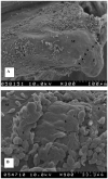
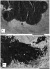
Scanning electron microscopy of the Randall's plaque. A. Randall's plaque appears as a protrusion on the papilla. Papillary surface epithelium (E) is sloughing exposing the sub-epithelial surface (SE). Dotted Bar = 100μm B. Higher magnification of an area in A showing surface epithelium (E), sub- epithelial surface (SE), as well as area of exposed fibrous layer (F) underneath. Dotted Bar = 33.3 μm C. Higher magnification of the fibrous layer. Dotted Bar = 20μm D. Spherulites of various diameters under the fibrous layer. Fibers as well as amorphous substances are also present. Dotted Bar = 15μm
Figure 2
Figure 2
Scanning electron microscopy of the Randall's plaque. A. Randall's plaque appears as a protrusion on the papilla. Papillary surface epithelium (E) is sloughing exposing the sub-epithelial surface (SE). Dotted Bar = 100μm B. Higher magnification of an area in A showing surface epithelium (E), sub- epithelial surface (SE), as well as area of exposed fibrous layer (F) underneath. Dotted Bar = 33.3 μm C. Higher magnification of the fibrous layer. Dotted Bar = 20μm D. Spherulites of various diameters under the fibrous layer. Fibers as well as amorphous substances are also present. Dotted Bar = 15μm
Figure 3
Figure 3
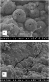
A. Higher magnification of the spherulites (SP) showing needle shaped crystallites on the surface as well as fibers (arrows) embedded therein. Dotted Bar = 4.29μm B. Surface of an internal layer of fused spehrulites. Dotted Bar = 30μm
Figure 4
Figure 4
Energy dispersive x-ray microanalysis of A. spherulites, and B. the fibers connecting them.
Figure 5
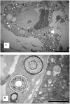
Figure 5. Transmission electron microscopic features of the Randall's plaque
A. A necrotic tubule is surrounded by laminated spherulites (arrows). They range in diameter from smaller than half a micron to approx 5 micron in diameter. Bar = 5μm B. Higher magnification of the spherulites with distinct concentric laminations and radial striations, present in an amorphous matrix under the renal tubular epithelium. Bar = 1μm
Figure 6
Figure 6
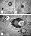
Higher magnification showing vesicular structures in and around basement membrane of the renal papillary collecting ducts. A. Small (arrowheads) and large vesicles (arrows) are dispersed in the basement membrane. Bar = 500nm B. A membrane (arrow) bound vesicle. Dotted Bar = 200nm C. Basement membrane of a collecting duct showing spherulites in grazinmg section. Surface appears uneven with projecting crystallites (arrows, compare with SEM appearance of the surface in Figure 3A). Collagen fiber (C) with banding pattern is also visible. Bar = 500nm
Figure 7
Figure 7
Transmission electron microscopic appearance of calcified deposits in the renal interstitium. A. Edge of the deposit demonstrating fusing spehrulites (arrows, compare with SEM appearance in Figure 3B). Some vesicular entities are also visible. Bar = 500nm B. Another area of large calcified deposit showing longitudinally running strands (ST). The deposit is surrounded by a matrix with collagen fibers (C). Bar = 5μm C. Fusion of spherical units (SP) of various dimensions with each other as well as calcified collagen produced a large calcified deposit surrounded by collagen fibers (C). Laminations can still be seen inside the peripheral spherulites (SP). Arrows point to the basement membrane of a collecting duct. Bar = 2μm D. Higher magnification of an area on the periphery of a large calcified deposit showing longitudinally running strands (ST). Notice the banding pattern (arrows) in the calcified strands. Compare it with the banding pattern in the nearby collagen fiber (C). Bar = 1μm
Figure 7
Figure 7
Transmission electron microscopic appearance of calcified deposits in the renal interstitium. A. Edge of the deposit demonstrating fusing spehrulites (arrows, compare with SEM appearance in Figure 3B). Some vesicular entities are also visible. Bar = 500nm B. Another area of large calcified deposit showing longitudinally running strands (ST). The deposit is surrounded by a matrix with collagen fibers (C). Bar = 5μm C. Fusion of spherical units (SP) of various dimensions with each other as well as calcified collagen produced a large calcified deposit surrounded by collagen fibers (C). Laminations can still be seen inside the peripheral spherulites (SP). Arrows point to the basement membrane of a collecting duct. Bar = 2μm D. Higher magnification of an area on the periphery of a large calcified deposit showing longitudinally running strands (ST). Notice the banding pattern (arrows) in the calcified strands. Compare it with the banding pattern in the nearby collagen fiber (C). Bar = 1μm
Figure 8
Figure 8
Selected area electron diffraction analyses at various sites on the calcified deposits showed different levels of crystallinity with A being mostly crystalline to C being near amorphous.

References

    1. Randall A. The Origin and Growth of Renal Calculi. Ann Surg. 1937;105:1009. - PMC - PubMed
    1. Coe FL, Evan AP, Worcester EM, et al. Three pathways for human kidney stone formation. Urol Res. 2010;38:147. - PMC - PubMed
    1. Evan AP, Lingeman JE, Coe FL, et al. Randall's plaque of patients with nephrolithiasis begins in basement membranes of thin loops of Henle. J Clin Invest. 2003;111:607. - PMC - PubMed
    1. Gower LB, Amos FF, Khan SR. Mineralogical signatures of stone formation mechanisms. Urol Res. 2010;38:281. - PMC - PubMed
    1. Murshed M, McKee MD. Molecular determinants of extracellular matrix mineralization in bone and blood vessels. Curr Opin Nephrol Hypertens. 19:359. - PubMed

Publication types