

Measuring the mechanical properties of blood clots formed via the tissue factor pathway of coagulation
- PMID: 22266209
- PMCID: PMC3778664
- DOI: 10.1016/j.ab.2011.12.036
Measuring the mechanical properties of blood clots formed via the tissue factor pathway of coagulation
Abstract
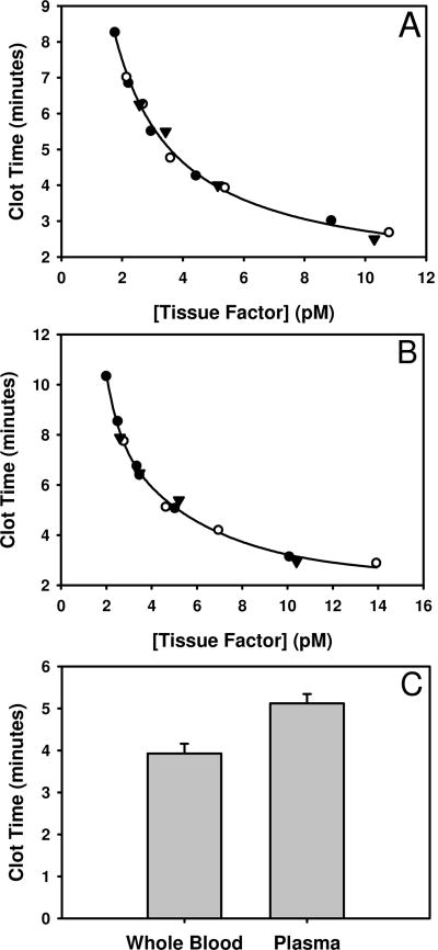
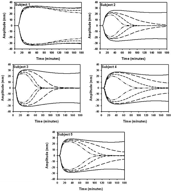
Thrombelastography (TEG) is a method that is used to conduct global assays that monitor fibrin formation and fibrinolysis and platelet aggregation in whole blood. The purpose of this study was to use a well-characterized tissue factor (Tf) reagent and contact pathway inhibitor (corn trypsin inhibitor, CTI) to develop a reproducible thrombelastography assay. In this study, blood was collected from 5 male subjects (three times). Clot formation was initiated in whole blood with 5 pM Tf in the presence of CTI, and fibrinolysis was induced by adding tissue plasminogen activator (tPA). Changes in viscoelasticity were then monitored by TEG. In quality control assays, our Tf reagent, when used at 5 pM, induced coagulation in whole blood in 3.93 ± 0.23 min and in plasma in 5.12 ± 0.23 min (n=3). In TEG assays, tPA significantly decreased clot strength (maximum amplitude, MA) in all individuals but had no effect on clot time (R time). The intraassay variability (CVa<10%) for R time, angle, and MA suggests that these parameters reliably describe the dynamics of fibrin formation and degradation in whole blood. Our Tf reagent reproducibly induces coagulation, making it an ideal tool to quantify the processes that contribute to mechanical clot strength in whole blood.
Copyright © 2012 Elsevier Inc. All rights reserved.
Figures
Similar articles
-
Evaluation of a modified thrombelastography assay initiated with recombinant human tissue factor in clinically healthy horses.Vet Clin Pathol. 2009 Dec;38(4):462-6. doi: 10.1111/j.1939-165X.2009.00157.x. Epub 2009 Jun 22. Vet Clin Pathol. 2009. PMID: 19548973
-
Comparison of coagulation and fibrinolysis in Irish Wolfhounds and age-matched control dogs using tissue plasminogen activator-augmented viscoelastic testing.J Vet Emerg Crit Care (San Antonio). 2024 May-Jun;34(3):222-230. doi: 10.1111/vec.13385. Epub 2024 May 22. J Vet Emerg Crit Care (San Antonio). 2024. PMID: 38775137
-
Thrombelastography in horses with acute gastrointestinal disease.J Vet Intern Med. 2011 Mar-Apr;25(2):307-14. doi: 10.1111/j.1939-1676.2010.0673.x. Epub 2011 Feb 11. J Vet Intern Med. 2011. PMID: 21314719
-
Global assays of fibrinolysis.Int J Lab Hematol. 2017 Oct;39(5):441-447. doi: 10.1111/ijlh.12688. Epub 2017 May 12. Int J Lab Hematol. 2017. PMID: 28497494 Review.
-
Viscoelasticity and Ultrastructure in Coagulation and Inflammation: Two Diverse Techniques, One Conclusion.Inflammation. 2015 Aug;38(4):1707-26. doi: 10.1007/s10753-015-0148-7. Inflammation. 2015. PMID: 25772112 Review.
Cited by
-
Thromboelastography and thrombin generation assessments for pediatric severe hemophilia A patients are highly variable and not predictive of clinical phenotypes.Res Pract Thromb Haemost. 2022 Sep 26;6(6):e12800. doi: 10.1002/rth2.12800. eCollection 2022 Aug. Res Pract Thromb Haemost. 2022. PMID: 36186102 Free PMC article.
-
Effect of BAX499 aptamer on tissue factor pathway inhibitor function and thrombin generation in models of hemophilia.Thromb Res. 2012 Dec;130(6):948-55. doi: 10.1016/j.thromres.2012.08.299. Epub 2012 Aug 27. Thromb Res. 2012. PMID: 22951415 Free PMC article.
-
Molecular dynamics simulations indicate that deoxyhemoglobin, oxyhemoglobin, carboxyhemoglobin, and glycated hemoglobin under compression and shear exhibit an anisotropic mechanical behavior.J Biomol Struct Dyn. 2018 May;36(6):1417-1429. doi: 10.1080/07391102.2017.1323674. Epub 2017 May 22. J Biomol Struct Dyn. 2018. PMID: 28441918 Free PMC article.
-
Global assays of hemostasis.Curr Opin Hematol. 2014 Sep;21(5):395-403. doi: 10.1097/MOH.0000000000000074. Curr Opin Hematol. 2014. PMID: 25054908 Free PMC article. Review.
-
Platelets do not express the oxidized or reduced forms of tissue factor.Biochim Biophys Acta. 2014 Mar;1840(3):1188-93. doi: 10.1016/j.bbagen.2013.11.024. Epub 2013 Dec 19. Biochim Biophys Acta. 2014. PMID: 24361609 Free PMC article.
References
-
- Nemerson Y. Tissue Factor And Hemostasis. Blood. 1988;71:1–8. - PubMed
-
- Gailani D, Renne T. The intrinsic pathway of coagulation: a target for treating thromboembolic disease? J Thromb Haemost. 2007;5:1106–1112. - PubMed
-
- Lapecorella M, Mariani G. Factor VII deficiency: defining the clinical picture and optimizing therapeutic options. Haemophilia. 2008;14:1170–1175. - PubMed
-
- Komiyama Y, Pedersen AH, Kisiel W. Proteolytic Activation of Human Factors IX and X by Recombinant Human Factor VIIa: Effects of Calcium, Phospholipids, and Tissue Factor. Biochemistry. 1990;29:9418–9425. - PubMed
Publication types
MeSH terms
Substances
Grants and funding
LinkOut - more resources
Full Text Sources
Miscellaneous
