. 2012 Mar 15;72(6):1557-67.
doi: 10.1158/0008-5472.CAN-11-3596. Epub 2012 Jan 20.

Kras(G12D) and p53 mutation cause primary intrahepatic cholangiocarcinoma

Affiliations

Kras(G12D) and p53 mutation cause primary intrahepatic cholangiocarcinoma

Michael R O'Dell et al. Cancer Res. .

Abstract

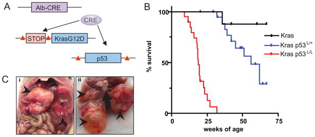
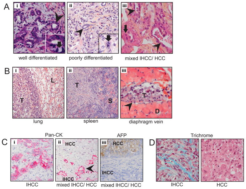
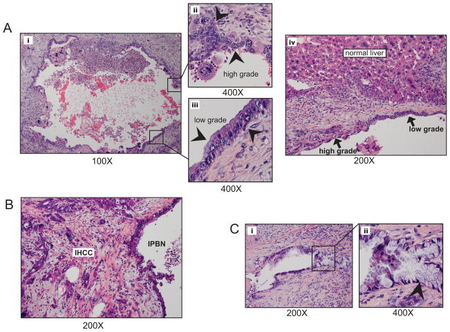
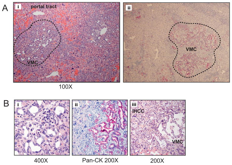
Intrahepatic cholangiocarcinoma (IHCC) is a primary cancer of the liver with an increasing incidence and poor prognosis. Preclinical studies of the etiology and treatment of this disease are hampered by the relatively small number of available IHCC cell lines or genetically faithful animal models. Here we report the development of a genetically engineered mouse model of IHCC that incorporates two of the most common mutations in human IHCC, activating mutations of Kras (Kras(G12D)) and deletion of p53. Tissue-specific activation of Kras(G12D) alone resulted in the development of invasive IHCC with low penetrance and long latency. Latency was shortened by combining Kras(G12D) activation with heterozygous or homozygous deletion of p53 (mean survival of 56 weeks vs. 19 weeks, respectively), which also resulted in widespread local and distant metastasis. Serial analysis showed that the murine models closely recapitulated the multistage histopathologic progression of the human disease, including the development of stroma-rich tumors and the premalignant biliary lesions, intraductal papillary biliary neoplasms (IPBN), and Von Meyenburg complexes (VMC; also known as biliary hamartomas). These findings establish a new genetically and histopathologically faithful model of IHCC and lend experimental support to the hypothesis that IPBN and VMC are precursors to invasive cancers.


Figures

Figure 1
Figure 1
KrasG12D and p53 mutation in the hepatic epithelium cooperate to cause cholangiocarcinoma. A) Modeling strategy; compound mutant mice harboring albumin-Cre transgene, LSL-KrasG12D, and p53L/L were created to conditionally activate KrasG12D and delete p53 in the hepatic epithelium. Lox P sites are indicated with red triangles. B) Kaplan-Meier survival analysis of cohorts of compound mutant animals. The p53 status is indicated; all of the mice have the LSL–KrasG12D allele. C) Gross tumor nodules in the Kras-p53 model. Arrowheads indicate tumor nodules seen in an animal with three distinct tumors. The arrow in image ii indicates enlarged lymph nodes. The tumor in the lower aspect of image ii is cystic and fluid- filled and was found to be an IPBN.
Figure 2
Figure 2
Kras-p53 IHCC recapitulates the histology of the human disease. A) Histology of primary liver tumors; from left to right i) well-differentiated IHCC 200X with inset demonstrating mucin producing cells (arrow), ii) poorly differentiated IHCC 200X with inset demonstrating mitotic figure (arrow) and, iii) mixed IHCC/HCC with arrow pointing towards area of HCC vs. glandular appearance of IHCC (arrowhead). B) Extensive metastasis (indicated by letter T) were observed grossly and histologically in the i) lung (L) ii) spleen (S) and iii) within the diaphragmatic lymphatic system. C) Pan-cytokeratin IHC of IHCC (i) and mixed IHCC/HCC (ii), upper region is non-staining HCC component and lower region showing glandular structures (arrowhead) which are pan-CK positive. AFP IHC of mixed IHCC/HCC (iii), upper region is AFP staining HCC component and lower region showing non-staining IHCC component. D) Trichrome IHC demonstrating collagen deposition within IHCC (left) but not in HCC (right).
Figure 3
Figure 3
IPBN is identified separately and in association with IHCC, demonstrating areas of low and high-grade cellular atypia. A) Fluid filled cystic structure (i; low power) demonstrated progressive cellular atypia (ii and iii) arrowheads point to epithelium showing an atypical low grade columnar biliary epithelium (ii) and high grade region with large nuclei, prominent nucleoli and marked cellular atypia (iii). IPBN were found within the context of normal liver (A;iv). B) IPBN adjacent and contiguous with invasive IHCC. C) Smaller IPBN (i) displayed an intestinal type epithelium (ii; arrowhead indicates mucin producing cell).
Figure 4
Figure 4
Von Meyenburg complexes (VCM), or biliary hamartomas, are prevalent and found in association with IHCC in the Kras-p53 model. A) i; Low-power view of Von Meyenburg Complex (circled in dashed black line between portal tracts). ii; IHC with pan-CK Ab of VMC within the hepatic parenchyma. B) i; VMC at high power demonstrated small caliber biliary epithelium surrounded by stroma, ii; VMC demonstrates strong staining with pan-CK Ab, iii; VMC associated with a well differentiated cholangiocarcinoma.
Figure 5
Figure 5
IHCC harbors molecular derangements characteristic of the human disease and activation of downstream RAS effecter pathways. A) Early passage (P1–3) IHCC cell lysates were tested for the expression of tumor suppressor genes. 25 μg of protein were loaded in each lane and murine pancreatic cell lines with previously defined genetic profiles (p53 or Ink4a/Arf mutant vs. wild-type) were used as controls (49). p53 expression is absent in all IHCC as is p21 expression. While the majority of tumors retained expression of p19Arf line #335 lost expression. Lines #460 and 335 both demonstrated absent p16Ink4a expression. SMAD4 expression was maintained across all lines. B) PCR reactions to detect the p53-WT (+) and p53lox alleles (Upper) and p53-null (−) allele (Lower) in normal tissue (tail clip) from p53lox/lox (lane 10), p53+/+ (lane 9), and p53 lox/+ (lane 8) mice and in tumor cell lines (lanes1–7). Primers and conditions used are previously described in detail (27). 4/6 tumor cell lines from Kras-p53L/+ animals (232, 338, 344, & 408) demonstrated no WT allele indicating loss of heterozygosity at the p53 locus. Tumor line 408 demonstrates retention of the p53-null allele with longer exposure (data not shown). Tumor cell line #449 from a Kras-p53+/+ animal shows retention of the p53+ allele. C) Western blot analysis of whole liver lysates from 10 weeks old wild-type and Kras-p53L/L animals compared with IHCC tumor lysates evaluating the relative activity of RAS effecters ERK and AKT demonstrating activity of ERK and AKT. Total LC3 levels were increased in IHCC as compared with normal and mutant liver (LC3 is lower band, upper band is non-specific).
Figure 6
Figure 6
Kras-p53 IHCC exhibits high basal levels of autophagy and is growth inhibited by chloroquine. A) IHCC cells were infected with a retrovirus expressing GFP-LC3 and grown in complete media with serum and fixed for confocal fluorescence microscopy. These were analyzed for the presence of LC3 dots, representing autophagic vesicles, and quantified. The % of autophagic cells (out of total number of 100 counted) with > 5 foci are shown in the upper right corners of each picture. Below is shown a western blot demonstrating high levels of LC3-II as compared with LC3-I, also consistent with a high level of basal autophagy. B) IHCC cells were cultured under normal growth conditions where the total number of LC3 foci was markedly increased in the presence of CQ (error bars are shown, students t-test is used for comparison, p< 0.001 for all cell lines treated). C & D) Growth of IHCC cell lines is inhibited in the presence of CQ p= <0.001 for samples 335, 518, and 339 and p= <0.01 for samples 449 (two-way ANOVA, error bars are shown for all data points, those not evident on the graphs fall within the area encompassed by the triangle or circle data-point). Consistent with a block in autophagic flux induced by CQ treatment we observed that treated lines had an accumulation of LC3-II and induction of p62 as compared with Day 0 control, with the exception of #518 where p62 expression was absent.

Comment in

References

    1. Hezel AF, Deshpande V, Zhu AX. Genetics of biliary tract cancers and emerging targeted therapies. J Clin Oncol. 2011;28:3531–40. - PMC - PubMed
    1. Shaib Y, El-Serag HB. The epidemiology of cholangiocarcinoma. Semin Liver Dis. 2004;24:115–25. - PubMed
    1. Valle J, Wasan H, Palmer DH, et al. Cisplatin plus gemcitabine versus gemcitabine for biliary tract cancer. N Engl J Med. 2011;362:1273–81. - PubMed
    1. Tannapfel A, Benicke M, Katalinic A, et al. Frequency of p16(INK4A) alterations and K-ras mutations in intrahepatic cholangiocarcinoma of the liver. Gut. 2000;47:721–7. - PMC - PubMed
    1. Tannapfel A, Sommerer F, Benicke M, et al. Mutations of the BRAF gene in cholangiocarcinoma but not in hepatocellular carcinoma. Gut. 2003;52:706–12. - PMC - PubMed

Publication types

Substances