

Factors affecting the endothelial retention of targeted microbubbles: influence of microbubble shell design and cell surface projection of the endothelial target molecule
- PMID: 22266330
- PMCID: PMC3941177
- DOI: 10.1016/j.echo.2011.12.016
Factors affecting the endothelial retention of targeted microbubbles: influence of microbubble shell design and cell surface projection of the endothelial target molecule
Abstract
Background: In biologic systems, the arrest of circulating cells is mediated by adhesion molecules projecting their active binding domain above the cell surface to enhance bond formation and tether strength. Similarly, molecular spacers are used for ligands on particle-based molecular imaging agents. The aim of this study was to evaluate the influence of tether length for targeting ligands on ultrasound molecular imaging agents.
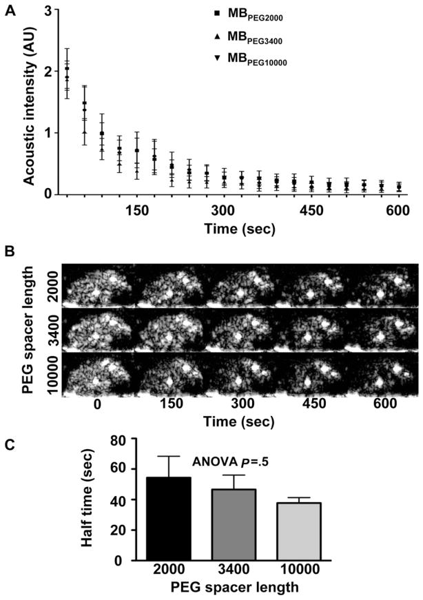
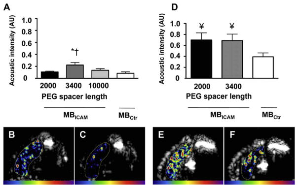
Methods: Microbubbles bearing biotin at the end of variable-length polyethylene glycol spacer arms (MB(2000) and MB(3400)) were prepared. To assess in vivo attachment efficiency to endothelial counterligands that vary in their distance from the endothelial cell surface, contrast-enhanced ultrasound (CEU) molecular imaging of tumor necrosis factor-α-induced P-selectin (long distance) or intercellular adhesion molecule-2 (short distance) was performed with each agent in murine hind limbs. To assess the influence of the glycocalyx on microbubble attachment, CEU molecular imaging of intercellular adhesion molecule-2 was performed after degradation of the glycocalyx.
Results: CEU molecular imaging targeted to P-selectin showed signal enhancement above control agent for MB(2000) and MB(3400), the degree of which was significantly higher for MB(3400) compared with MB(2000). CEU molecular imaging targeted to intercellular adhesion molecule-2 showed low overall signal for all agents and signal enhancement above control for MB(3400) only. Glycocalyx degradation increased signal for MB(3400) and MB(2000).
Conclusions: Microbubble targeting to endothelial ligands is influenced by (1) the tether length of the ligand, (2) the degree to which the endothelial target is projected from the cell surface, and (3) the status of the glycocalyx. These considerations are important for designing targeted imaging probes and understanding potential obstacles to molecular imaging.
Copyright © 2012 American Society of Echocardiography. Published by Mosby, Inc. All rights reserved.
Figures


References
-
- Kaufmann BA, Lindner JR. Molecular imaging with targeted contrast ultrasound. Curr Opin Biotechnol. 2007;18:11–6. - PubMed
-
- Kaufmann BA, Lewis C, Xie A, Mirza-Mohd A, Lindner JR. Detection of recent myocardial ischaemia by molecular imaging of P-selectin with targeted contrast echocardiography. Eur Heart J. 2007;28:2011–7. - PubMed
-
- Behm CZ, Kaufmann BA, Carr C, Lankford M, Sanders JM, Rose CE, et al. Molecular imaging of endothelial vascular cell adhesion molecule-1 expression and inflammatory cell recruitment during vasculogenesis and ischemia-mediated arteriogenesis. Circulation. 2008;117:2902–11. - PubMed
-
- Kaufmann BA. Ultrasound molecular imaging of atherosclerosis. Cardiovasc Res. 2009;83:617–25. - PubMed
Publication types
MeSH terms
Substances
Grants and funding
LinkOut - more resources
Full Text Sources
Miscellaneous
