Review
. 2012 Mar;33(3):300-11.
doi: 10.1038/aps.2011.170. Epub 2012 Jan 23.

Structure and mechanism for recognition of peptide hormones by Class B G-protein-coupled receptors

Affiliations
Review

Structure and mechanism for recognition of peptide hormones by Class B G-protein-coupled receptors

Kuntal Pal et al. Acta Pharmacol Sin. 2012 Mar.

Abstract

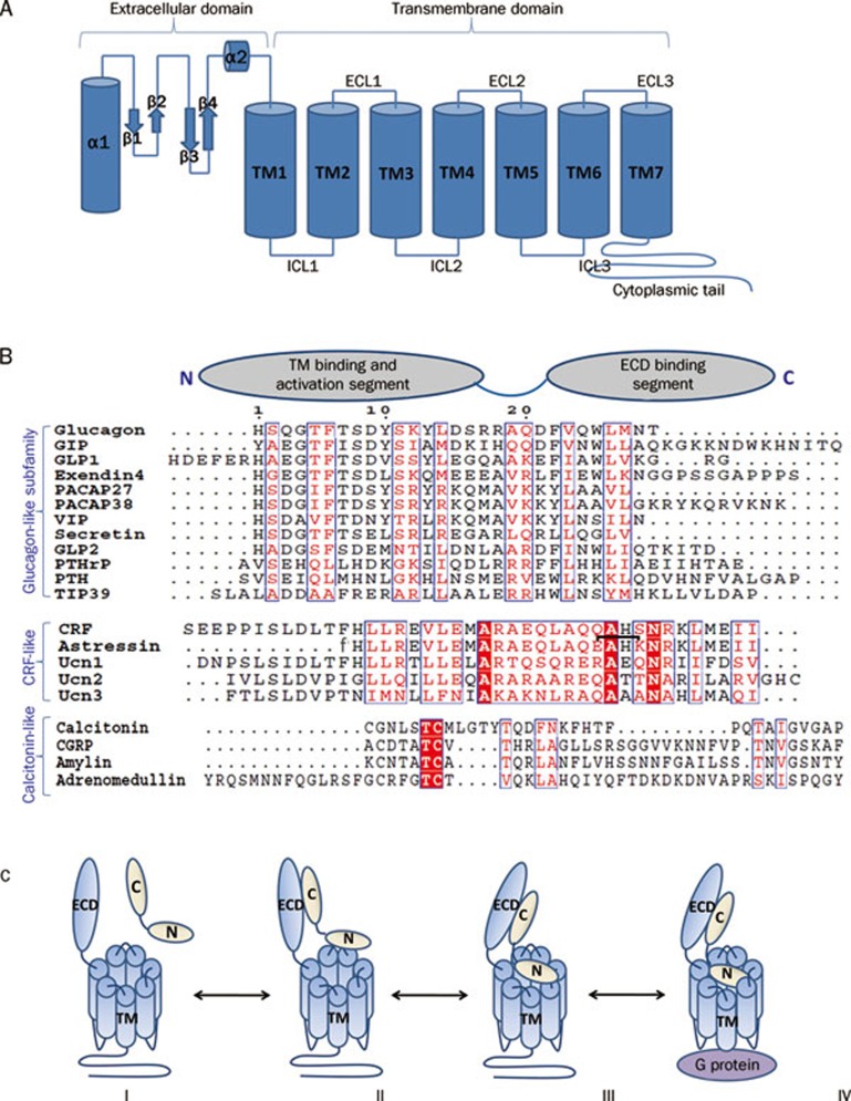
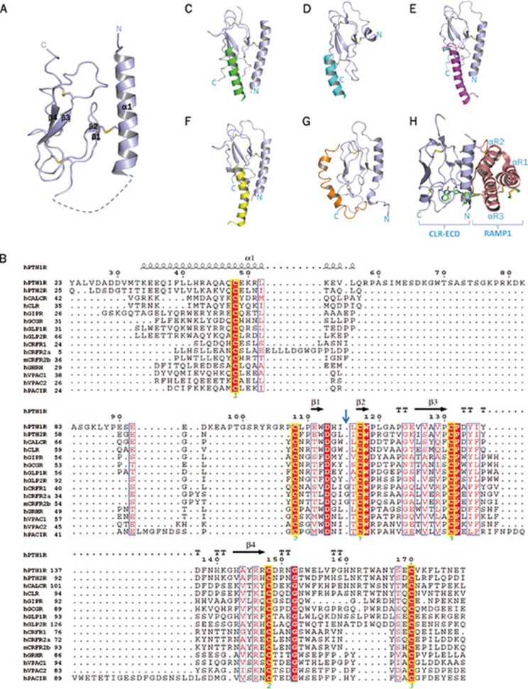
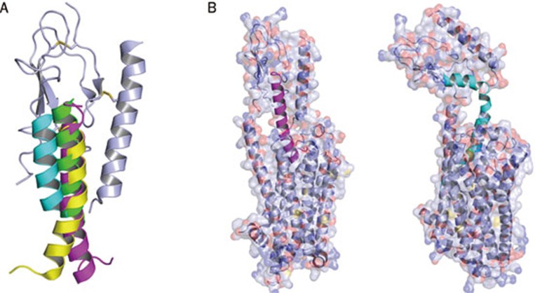
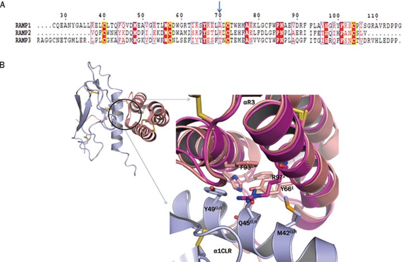
Class B G-protein-coupled receptors (GPCRs) are receptors for peptide hormones that include glucagon, parathyroid hormone, and calcitonin. These receptors are involved in a wide spectrum of physiological activities, from metabolic regulation and stress control to development and maintenance of the skeletal system. As such, they are important drug targets for the treatment of diabetes, osteoporosis, and stress related disorders. Class B GPCRs are organized into two modular domains: an extracellular domain (ECD) and a helical bundle that contains seven transmembrane helices (TM domain). The ECD is responsible for the high affinity and specificity of hormone binding, and the TM domain is required for receptor activation and signal coupling to downstream G-proteins. Although the structure of the full-length receptor remains unknown, the ECD structures have been well characterized for a number of Class B GPCRs, revealing a common fold for ligand recognition. This review summarizes the general structural principles that guide hormone binding by Class B ECDs and their implications in the design of peptide hormone analogs for therapeutic purposes.


Figures

Figure 1
Figure 1
(A) Cartoon presentation of the general architecture of Class B GPCRs consisting of a N-terminal extracellular domain (ECD) and a C-terminal transmembrane domain (7TM). The ECD forms a three layer α-β-β/α fold and the 7TM domain seven membrane-spanning helices connected by three extracellular loops (ECLs) and three intracellular loops (ICLs). (B) Sequence alignment of Class B GPCR ligands with cartoon presentation of their N- and C-terminal domains on top. Based on sequence similarity, ligands can be grouped into glucagon-like, CRF-like, and calcitonin-like subfamilies. Identical residues are shown as white letters on red background. Partially conserved residues are shown as red letters. The residue numbering on top corresponds to that of glucagon. The lactam bridge in astressin is indicated by a black bracket, “f” in the astressin sequence indicates D-phenylalanine. (C) Two domain binding model for class B GPCRs. (I) Peptide hormone and receptor are orientated for initial receptor ligand binding. (II) The initial complex forms between the C-terminus of the peptide and the ECD of the receptor. (III) This interaction facilitates the binding of the free N-terminus of the peptide to the juxtamembrane region of the 7TM domain of the receptor. (IV) This binding induces a conformational change in the 7TM and cytoplasmic domain of the receptor, which mediates its interaction with a heterotrimeric G protein.
Figure 2
Figure 2
(A) A ribbon diagram of the basic architecture of the “secretin family recognition fold” of the extracellular domain of class B GPCRs. The structure is mainly divided into three layers consisting of an N terminal α helix and two pairs of antiparallel β sheets. The conserved disulfide bonds connecting the three layers are depicted as sticks. (B) Sequence alignment of the extracellular domains of human Class B GPCRs with secondary structure elements for PTH1R indicated on top (PDB: 3C4M). Invariant and conserved residues are highlighted. The glycine residues specific for the CRFR subfamily are marked by a blue arrow. Invariant cysteine residues are indicated by a yellow box. Identical residues are shown as white letters on red background. Partially conserved residues are shown as red letters on white background. The residue numbering on top corresponds to that of hPTH1R. Cysteine pairs forming disulfide bonds are indicated by yellow outlines and by green numbers at the bottom. TT=tight turns. (C) Structure of the hPTH1R-PTH ECD complex with the ECD shown in light blue and PTH in green. (D) Structure of the hCRFR1-CRF complex with the ECD shown in light blue and CRF in cyan. (E) Structure of the hGIPR-GIP complex with the ECD in light blue and GIP in magenta. (F) Structure of the hGLP1R-Glp1 complex with the ECD in light blue and Glp1 in yellow. (G) Structure of the PAC1R-PACAP complex with the ECD in light blue and PACAP in orange. (H) Structure of the CLR-Telcagepant (a small molecule drug for the treatment of migraine) with the CLR-ECD in light blue and RAMP1 in salmon.
Figure 3
Figure 3
(A) Structural alignment of ECD-bound Class B GPCR ligands. The ligands form helical conformations with their C termini interacting with the ECD. The N termini remain free and show a high level of flexibility. The ligands shown are PTH(15–34) in green, GIP(1–42) in magenta, GLP1(7–37) in yellow, and CRF(22–41) in cyan. (B) Models displaying possible hormone positions in the context of full length receptors. Models of ECD-bound GIP(1–42) (magenta) [PDB: 2QKH] and a modified CRF (cyan) [PDB: 2L27] were superpositioned on a model of the transmembrane domain of turkey β1-adrenergic receptor [PDB: 2Y03]. The ECD was adjusted manually with distance constraints using COOT. The different binding positions between CRF-like and glucagon-like subfamily peptides suggest that the ECDs may also adopt two different conformations in the context of full length receptors. Note that these models just illustrate relative dimensions of receptors and ligands as well as predictions of ligand binding sites. Only structures of the complexes between full length receptors and their ligands can provide accurate position and conformation of receptor-bound ligands.
Figure 4
Figure 4
(A) Sequence alignment of RAMP 1, 2, and 3. The important non-conserved residue in the RAMP1-CLR interaction pocket has been highlighted by a blue arrow. Invariant cysteine residues have been shown in yellow box. Identical residues are shown as white letters on red background. Partially conserved residues are shown as red letters. The residue numbering on top corresponds to that of RAMP1. (B) Structural representation of the CGRP receptor with the CLR-ECD in light blue and RAMP1 in salmon. Structural alignment of the binding interface of the RAMP1-CLR complex (salmon-lightblue; PDB: 3N7S) with apo RAMP2 (magenta; PDB: 2XVT). The helices 2 and 3 of RAMP have been marked. The side chains of RAMP2 R97 and CLR Q45CLR in the binding interface sterically clash.

References

    1. Fredriksson R, Lagerstrom MC, Lundin LG, Schioth HB. The G-protein-coupled receptors in the human genome form five main families. Phylogenetic analysis, paralogon groups, and fingerprints. Mol Pharmacol. 2003;63:1256–72. - PubMed
    1. Foord SM, Bonner TI, Neubig RR, Rosser EM, Pin JP, Davenport AP, et al. International Union of Pharmacology. XLVI. G protein-coupled receptor list. Pharmacol Rev. 2005;57:279–88. - PubMed
    1. Harmar AJ. Family-B G-protein-coupled receptors. Genome Biol. 2001;2:REVIEWS3013. - PMC - PubMed
    1. Lagerstrom MC, Schioth HB. Structural diversity of G protein-coupled receptors and significance for drug discovery. Nat Rev Drug Discov. 2008;7:339–57. - PubMed
    1. Grace CR, Perrin MH, DiGruccio MR, Miller CL, Rivier JE, Vale WW, et al. NMR structure and peptide hormone binding site of the first extracellular domain of a type B1 G protein-coupled receptor. Proc Natl Acad Sci U S A. 2004;101:12836–41. - PMC - PubMed

Publication types

MeSH terms