. 2012 Nov 8;31(45):4750-8.
doi: 10.1038/onc.2011.633. Epub 2012 Jan 23.

Scavenging of CXCL12 by CXCR7 promotes tumor growth and metastasis of CXCR4-positive breast cancer cells

Affiliations

Scavenging of CXCL12 by CXCR7 promotes tumor growth and metastasis of CXCR4-positive breast cancer cells

K E Luker et al. Oncogene. .

Abstract

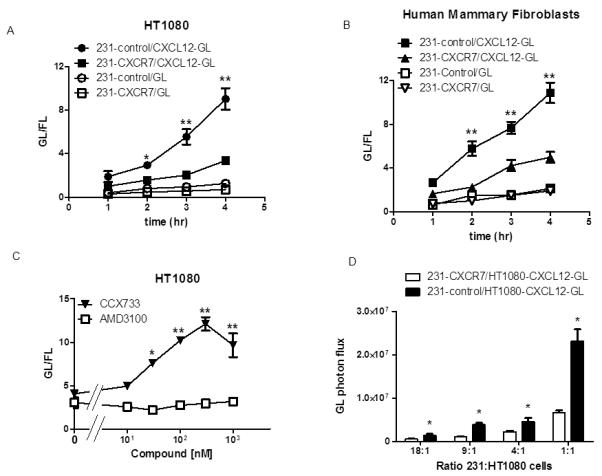
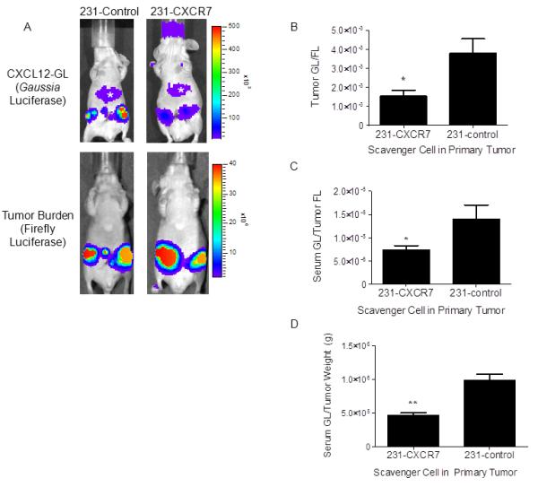
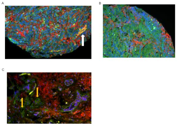
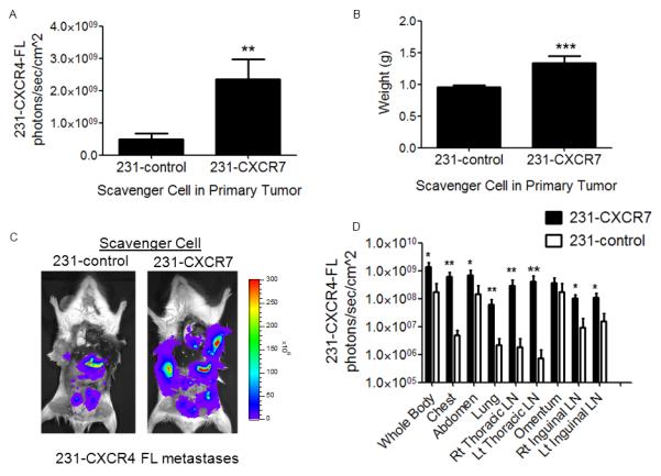
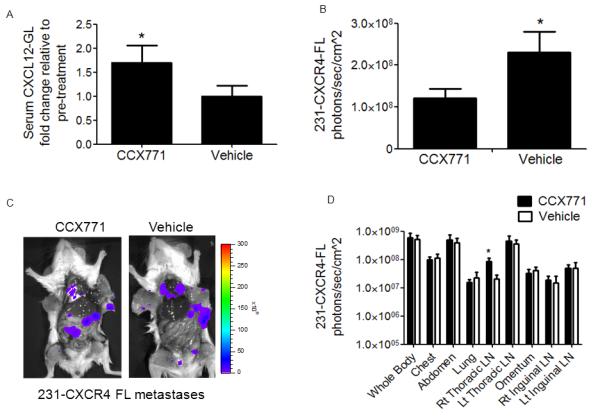
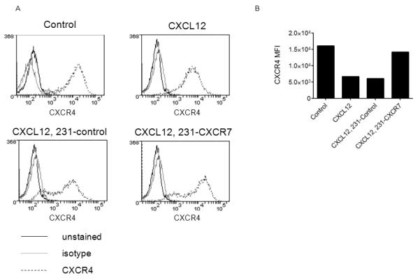
Chemokine CXCL12 and receptor CXCR4 control multiple steps in primary tumor growth and metastasis in breast cancer and more than 20 other human malignancies. Mechanisms that regulate availability of CXCL12 in tumor microenvironments will substantially impact cancer progression and ongoing efforts to target the CXCL12-CXCR4 pathway for cancer chemotherapy. We used dual luciferase imaging to investigate CXCR7-dependent scavenging of CXCL12 in breast tumors in vivo and quantify effects of CXCR7 on tumor growth and metastasis of a separate population of CXCR4+ breast cancer cells. In a mouse xenograft model of human breast cancer, in vivo imaging showed that malignant cells expressing CXCR7 reduced bioluminescent CXCL12 secreted in the primary tumor microenvironment. Capitalizing on sensitive detection of bioluminescent CXCL12, we also demonstrated that CXCR7+ cells reduced amounts of chemokine released from orthotopic tumors into the circulation. Immunofluorescence staining of human primary breast cancers showed expression of CXCR4 and CXCR7 on malignant cells in ≈30% of cases. In most cases, CXCR4 and CXCR7 predominantly were expressed on separate populations of malignant cells in a tumor. We modeled these cases of human breast cancer by co-implanting tumor xenografts with CXCR4+ breast cancer cells, human mammary fibroblasts secreting CXCL12, and CXCR7+ or control breast cancer cells. Bioluminescence imaging showed that CXCR7+ breast cancer cells enhanced proliferation of CXCR4+ breast cancer cells in orthotopic tumors and spontaneous metastases. Treatment with a small-molecule inhibitor of CXCR7 chemokine limited the growth of CXCR4+ breast cancer cells in tumors that also contained malignant CXCR7+ cells. These studies establish a new in vivo imaging method to quantify chemokine scavenging by CXCR7 in the tumor microenvironment and identify that CXCR7+ cells promote growth and metastasis of CXCR4+ breast cancer cells.


Figures

Figure 1
Figure 1. CXCR7 removes CXCL12 from the extracellular space
A) MD-MBA-231 breast cancer cells stably transduced with CXCR7 (231-CXCR7) or GFP (231-control) were co-cultured overnight with equal numbers of HT1080 cells secreting either CXCL12-GL or unfused GL, respectively. Amounts of bioluminescent CXCL12-GL and GL in the extracellular space were quantified at various times through 4 hours (n = 4 per condition). B) Co-cultures of 231-CXCR7 or 231-control cells with human mammary fibroblasts secreting either CXCL12-GL or unfused GL were analyzed as in A (n = 4 per condition). C) Co-cultures of 231-CXCR7 and HT1080-CXCL12-GL cells were treated with increasing concentrations of inhibitors of CXCR7 (CCX733) or CXCR4 (AMD3100) for 4 hours before quantifying amounts of CXCL12-GL in culture medium (n = 4 per condition). D) Various ratios of 231-CXCR7 or 231-control cells to HT1080-CXCL12-GL cells were incubated for 4 hours before quantifying CXCL12-GL in culture medium (n = 4 per condition). Data are presented as mean values + SEM. *, p < 0.05; **, p < 0.01.
Figure 2
Figure 2. CXCR7 reduces CXCL12 in primary breast tumors
A) Mice were implanted with orthotopic breast tumor xenografts of 231-CXCR7 or 231-control cells with HT1080-CXCL12-GL/FL cells. Representative images are presented from Gaussia and firefly luciferase bioluminescence imaging for tumors with 231-CXCR7 or 231-control cells. Asterisk denotes bioluminescence from oxidation of coelenterazine in liver. Scale bar depicts range of photon flux values as pseudocolor display with red and blue representing high and low values, respectively. B) Photon flux from CXCL12-GL was normalized to firefly luciferase in each tumor and presented as mean values + SEM (n = 5 per group). C) CXCL12-GL in serum was quantified by bioluminescence. Data were normalized to photon flux from firefly luciferase in each mouse. D) Bioluminescence from CXCL12-GL in serum was normalized to weight of excised tumors. Data were graphed as mean values + SEM. *, p < 0.05; **, p < 0.01.
Figure 3
Figure 3. CXCR4 and CXCR7 expression in primary human breast cancers
A-C) Tissue microarrays from primary human breast cancers were stained for CXCR4 (red) and CXCR7 (green). Nuclei were stained with DAPI (blue). Representative images from 3 different tumors are shown. A, B) CXCR4 and CXCR7 are present on separate populations of malignant cells, and white arrow in A shows malignant cells that co-express CXCR4 and CXCR7. C) CXCR4+ breast cancer cells are present with CXCR7 on endothelial cells in tumor blood vessels (orange arrows).
Figure 4
Figure 4. CXCR7 in the primary tumor microenvironment increases growth and metastasis of breast cancer cells expressing CXCR4
Mice were implanted with orthotopic breast tumor xenografts comprised of human mammary fibroblasts expressing CXCL12-GL (HMF-CXCL12-GL) and 231-CXCR4-FL cells combined with either 231-CXCR7 or 231-control cells (n = 10 per group). A) Mean values + SEM for firefly luciferase bioluminescence after 4 weeks of tumor growth from 231-CXCR4-FL cells in mice with either 231-control or 231-CXCR7 scavenger cells. B) Mean values for weights of excised tumors from each group of mice. C) Representative firefly luciferase bioluminescence images of metastases from 231-CXCR4-FL cells in a mouse from each group. Scale bar shows pseudocolor display for photon flux with red and blue representing highest and lowest values, respectively. D) Quantified data for 231-CXCR4-FL bioluminescence in metastatic sites for mice with either 231-control or 231-CXCR7 scavenger cells. Data were presented as mean values + SEM. *, p < 0.05; **, p < 0.01; ***, p < 0.005.
Figure 5
Figure 5. Inhibiting CXCR7 reduces growth of CXCR4+ breast cancer cells in orthotopic tumors
Mice were implanted with orthotopic breast tumor xenografts comprised of human mammary fibroblasts expressing CXCL12-GL (HMF-CXCL12-GL), 231-CXCR4-FL cells, and 231-CXCR7 cells (n = 10 per group). After palpable tumors developed one week after implantation, mice were treated daily with subcutaneous injections of CXCR7 inhibitor CCX771 (30 mg/kg) or vehicle control. A) CXCL12-GL in serum samples was quantified after one week of treatment. Data are presented as mean values + SEM. B) Quantified firefly luciferase bioluminescence from 231-CXCR4-FL cells in primary tumors after 4 weeks of tumor growth. Graph shows mean values + SEM. C) Representative firefly luciferase bioluminescence images of 231-CXCR4-FL metastases in mice treated with CCX771 or vehicle control. Scale bar depicts range of values for photon flux as a pseudocolor display. D) Quantified data for photon flux from 231-CXCR4-FL metastases in multiple anatomic sites. Graph displays mean values + SEM. *, p < 0.05.
Figure 6
Figure 6. CXCR7 regulates cell surface expression of CXCR4 and CXCL12-dependent signaling
A) Flow cytometry plots for expression of cell surface CXCR4 in 231-CXCR4 cells cultured for 2 days in serum-free medium alone (control), 100 ng/ml CXCL12 (CXCL12), 100 ng/ml CXCL12 plus co-cultured 231-control cells (CXCL12, 231-control), or 100 ng/ml CXCL12 plus co-cultured 231-CXCR7 cells (CXCL12, 231-CXCR7). Black line, unstained cells; gray line, isotype antibody; dashed line, CXCR4 antibody. B) Graph displays mean fluorescence intensity for cell surface CXCR4 from flow cytometry.

References

    1. Smith M, Luker K, Garbow J, Prior J, Jackson E, Piwnica-Worms D, et al. CXCR4 regulates growth of both primary and metastatic breast cancer. Cancer Res. 2004;64:8604–8612. - PubMed
    1. Orimo A, Gupta P, Sgroi D, Arenzana-Seisdedos F, Delaunay T, Naeem R, et al. Stromal fibroblasts present in invasive human breast carcinomas promote tumor growth and angiogenesis through elevated SDF-1/CXCL12 secretion. Cell. 2005;121:335–348. - PubMed
    1. Zou W, Machelon V, Coulomb-L’Hermin A, Borvak J, Nome F, Isaeva T, et al. Stromal-derived factor-1 in human tumors recruits and alters the function of plasmacytoid precursor dendritic cells. Nat Med. 2001;7:1339–1346. - PubMed
    1. Lee B, Lee T, Avraham S, Avraham H. Involvement of the chemokine receptor CXCR4 and its ligand stromal cell-derived factor 1alpha in breast cancer cell migration through human brain microvascular endothelial cells. Mol Cancer Res. 2004;2:327–338. - PubMed
    1. Muller A, Homey B, Soto H, Ge N, Catron D, Buchanon M, et al. Involvement of chemokine receptors in breast cancer metastasis. Nature. 2001;410:50–56. - PubMed

Publication types

MeSH terms