. 2012 Nov 15;31(46):4859-67.
doi: 10.1038/onc.2011.647. Epub 2012 Jan 23.

Antibody targeting of anaplastic lymphoma kinase induces cytotoxicity of human neuroblastoma

Affiliations

Antibody targeting of anaplastic lymphoma kinase induces cytotoxicity of human neuroblastoma

E L Carpenter et al. Oncogene. .

Erratum in

  • Oncogene. 2012 Nov 15;31(46):4888. Vigny, M [added]; Mazot, P [added]

Abstract

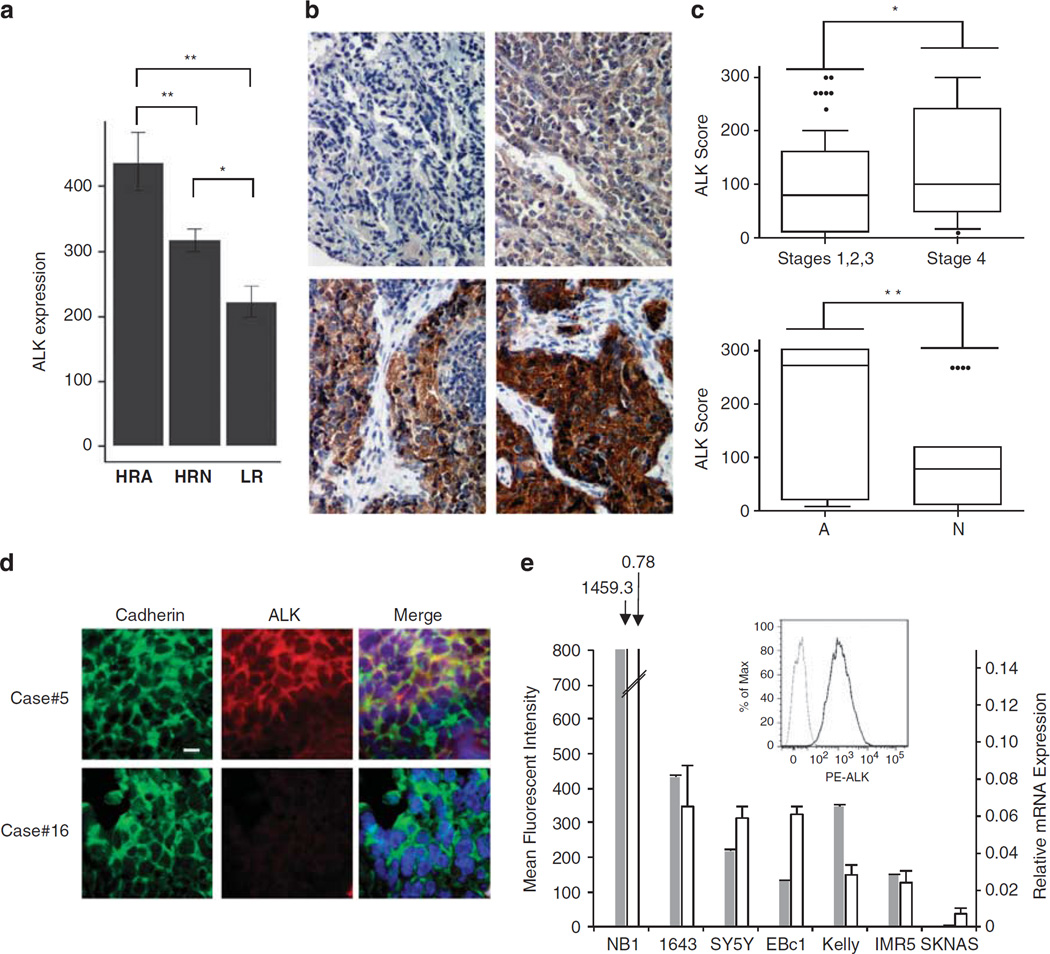
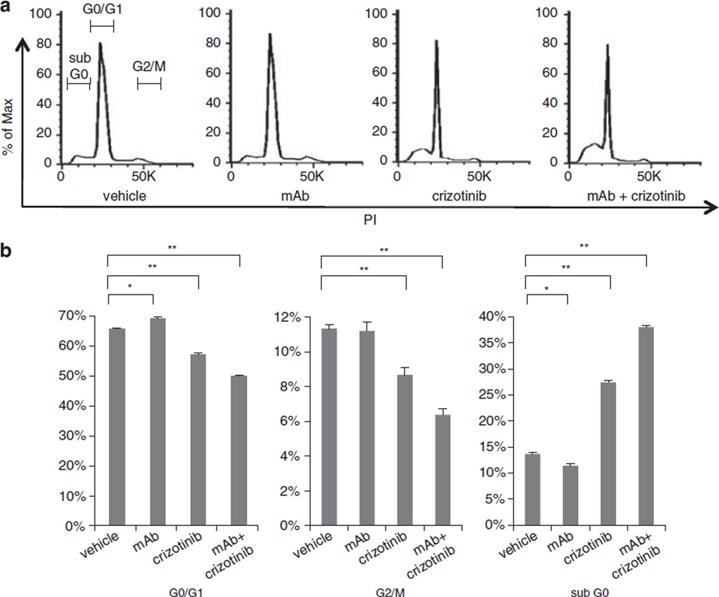
Anaplastic lymphoma kinase (ALK) is a receptor tyrosine kinase aberrantly expressed in neuroblastoma, a devastating pediatric cancer of the sympathetic nervous system. Germline and somatically acquired ALK aberrations induce increased autophosphorylation, constitutive ALK activation and increased downstream signaling. Thus, ALK is a tractable therapeutic target in neuroblastoma, likely to be susceptible to both small-molecule tyrosine kinase inhibitors and therapeutic antibodies-as has been shown for other receptor tyrosine kinases in malignancies such as breast and lung cancer. Small-molecule inhibitors of ALK are currently being studied in the clinic, but common ALK mutations in neuroblastoma appear to show de novo insensitivity, arguing that complementary therapeutic approaches must be developed. We therefore hypothesized that antibody targeting of ALK may be a relevant strategy for the majority of neuroblastoma patients likely to have ALK-positive tumors. We show here that an antagonistic ALK antibody inhibits cell growth and induces in vitro antibody-dependent cellular cytotoxicity of human neuroblastoma-derived cell lines. Cytotoxicity was induced in cell lines harboring either wild type or mutated forms of ALK. Treatment of neuroblastoma cells with the dual Met/ALK inhibitor crizotinib sensitized cells to antibody-induced growth inhibition by promoting cell surface accumulation of ALK and thus increasing the accessibility of antigen for antibody binding. These data support the concept of ALK-targeted immunotherapy as a highly promising therapeutic strategy for neuroblastomas with mutated or wild-type ALK.


Conflict of interest statement

CONFLICT OF INTEREST

The authors declare no conflict of interest.

Figures

Figure 1
Figure 1
ALK expression in neuroblastoma. (a) ALK expression in 229 neuroblastoma patient tumors analyzed by Affymetrix Human Exon Array and normalized using quantile normalization (high-risk MYCN amplified (HRA), n=64; high-risk MYCN non-amplified (HRN), n=141; low-risk (LR), n= 24). (b) Representative images for immunohistochemical staining of ALK in neuroblastoma patient tumors. ALK staining was positive overall in 109 of 126 (86.5%) samples analyzed. In all, 17 samples showed no positive staining (upper left panel); 35 showed weak staining (upper right panel); 55 showed moderate staining (lower left panel); and 19 showed strong staining (lower right panel). (c) Box plots showing 10th percentile, 90th percentile and mean ALK score by immunohistochemistry for INSS stage (top panel) and MYCN status (bottom panel; A, MYCN amplified; N, MYCN non-amplified). **P<0.01; *P<0.05. ****indicate data-points outside of percentiles. (d) Representative confocal micrographs of formalin-fixed paraffin-embedded primary neuroblastoma patient tumor slides showing staining for ALK and the cell surface antigen cadherin. All samples showed strong plasma membrane cadherin staining, and of 17 samples stained, 1 exhibited strong ALK staining (ALK score=3; top row), 10 showed intermediate ALK staining, 2 had weak staining and 3 had no ALK staining (ALK score=0, bottom row). Bar represents 10µm. (e) Comparison of flow cytometry results with ALK mRNA expression levels for several neuroblastoma cell lines. Gray bars show flow cytometry results for ALK cell surface staining. White bars represent an ALK mRNA expression ‘index’ (relative expression) measured as ALK levels relative to HPRT1. Inset shows mean fluorescence intensity of NB1 cells for ALK staining (black line) and isotype control (gray line).
Figure 2
Figure 2
ALK antibody-induced growth inhibition and ADCC of neuroblastoma cells. To measure growth inhibition upon antibody exposure, cell lines were plated in 96-well plates and treated with anti-ALK (mAb30 plus mAb49) or a negative control murine IgG1. (a) Growth inhibition of SY5Y cells treated with indicated amounts of anti-ALK as compared with control Ig. (b) The indicated cell lines were treated with 10 µg/ml ALK antibody, and growth inhibition was measured after 144h. (c) ADCC was measured using an in vitro assay as described in Materials and methods, in which IL-2-activated peripheral blood lymphocytes were co-incubated with neuroblastoma cells in the presence (black line) or absence (gray line) of 1 µg/ml ALK antibody. Shown are percent (%) cytotoxicities at the indicated effector:target ratios when NB1 cells (left panel), SY5Y cells (middle panel) or cell surface ALK-negative SKNAS cells (right panel) were used as targets.
Figure 3
Figure 3
Effect of crizotinib on cell surface ALK expression. (a) SY5Y cells were incubated for 24h with vehicle or 1000 nm crizotinib, then biotin labeled, precipitated with NeutrAvidin beads and immunoblotted for ALK (upper left panel) or cadherin (lower left panel). The results of densitometric analysis of ALK protein, normalized to cadherin, are shown in the bar chart (right panel), and indicate an increase in total ALK from 1.39 to 2.91 in arbitrary units. (b) Representative flow cytometry results showing mean fluorescence intensity (MFI) for SY5Y cells incubated for 72 h with either vehicle (medium gray line; MFI = 420) or 1000 nm crizotinib (black line; MFI= 636), resulting in a 51.4% increase in MFI for crizotinib- versus vehicle-treated cells. The single-peaked light gray line represents the isotype control. (c) Concentration dependence of the percent change in cell surface ALK, as measured by MFI, for cells incubated for 72 h with varying concentrations of crizotinib as compared with vehicle. (d) Time course of percentage change in cell surface ALK levels (over that seen for vehicle treatment) when cells were incubated with 1000 nm crizotinib.
Figure 4
Figure 4
Dual antibody/TKI targeting of ALK. SY5Y cells were treated with either 333 nm crizotinib or 10 µg/ml anti-ALK antibody (mAb30+mAb49) or both. As negative control, cells were treated with equal volumes of dimethyl sulfoxide and 10µg/ml murine IgG1. (a) Cell growth was monitored by Real-Time Cell Electronic Sensing. (b) Immunoblot analysis of native ALK protein levels (upper panel) and phospho-ALK (middle panel). β-Actin levels are shown as a loading control (lower panel). (c) Effect of crizotinib pretreatment on anti-ALK antibody-mediated ADCC was measured. SY5Y cells were preincubated in the presence of crizotinib or vehicle for 48h, harvested and then used as target cells in the in vitro ADCC assay. **P<0.01 and *P<0.05.
Figure 5
Figure 5
Effects of an antagonist ALK antibody on crizotinib dose - response curve. SY5Y cells were treated with crizotinib at the indicated doses, either alone or in combination with 10µg/ml (total) ALK antibody mAb30 and mAb49. Cell growth was measured at day 7 using Real-Time Cell Electronic Sensing. (a) Comparison of growth inhibition for monotherapy with dual ALK targeting: white bars represent crizotinib alone and gray bars represent crizotinib treatment in the presence of 10µg/ml anti-ALK. **P<0.01 and *P<0.05. (b) IC50 was calculated over a range of 10 doses of crizotinib alone (circles) or crizotinib plus 10 µg/ml anti-ALK (squares), yielding IC50 values of 3018 nm and 1745 nm, respectively.
Figure 6
Figure 6
Cell cycle analysis of inhibitor-treated cells. SY5Y cells were treated with 1000 nm crizotinib, 10 µg/ml antibody, both, or vehicle/IgG1, and were then harvested, fixed, stained with propidium iodide and analyzed by flow cytometry. (a) Representative histograms showing proportion of cells in sub-G0/apoptosis, G0/G1 and G2/mitosis. (b) Quantification of flow cytometry results. **P<0.01 and *P<0.05.

References

    1. Morris SW, Kirstein MN, Valentine MB, Dittmer KG, Shapiro DN, Saltman DL, et al. Fusion of a kinase gene, ALK, to a nucleolar protein gene, NPM, in non-Hodgkin’s lymphoma. Science. 1994;263:1281–1284. - PubMed
    1. Rikova K, Guo A, Zeng Q, Possemato A, Yu J, Haack H, et al. Global survey of phosphotyrosine signaling identifies oncogenic kinases in lung cancer. Cell. 2007;131:1190–1203. - PubMed
    1. Soda M, Choi YL, Enomoto M, Takada S, Yamashita Y, Ishikawa S, et al. Identification of the transforming EML4-ALK fusion gene in non-small-cell lung cancer. Nature. 2007;448:561–566. - PubMed
    1. Jazii FR, Najafi Z, Malekzadeh R, Conrads TP, Ziaee AA, Abnet C, et al. Identification of squamous cell carcinoma associated proteins by proteomics and loss of beta tropomyosin expression in esophageal cancer. World J Gastroenterol. 2006;12:7104–7112. - PMC - PubMed
    1. Griffin CA, Hawkins AL, Dvorak C, Henkle C, Ellingham T, Perlman EJ. Recurrent involvement of 2p23 in inflammatory myofibroblastic tumors. Cancer Res. 1999;59:2776–2780. - PubMed

Publication types

MeSH terms