. 2012 Jan 1;125(Pt 1):37-48.
doi: 10.1242/jcs.077040. Epub 2012 Jan 20.

TOR complex 2 (TORC2) in Dictyostelium suppresses phagocytic nutrient capture independently of TORC1-mediated nutrient sensing

Affiliations

TOR complex 2 (TORC2) in Dictyostelium suppresses phagocytic nutrient capture independently of TORC1-mediated nutrient sensing

Daniel Rosel et al. J Cell Sci. .

Abstract

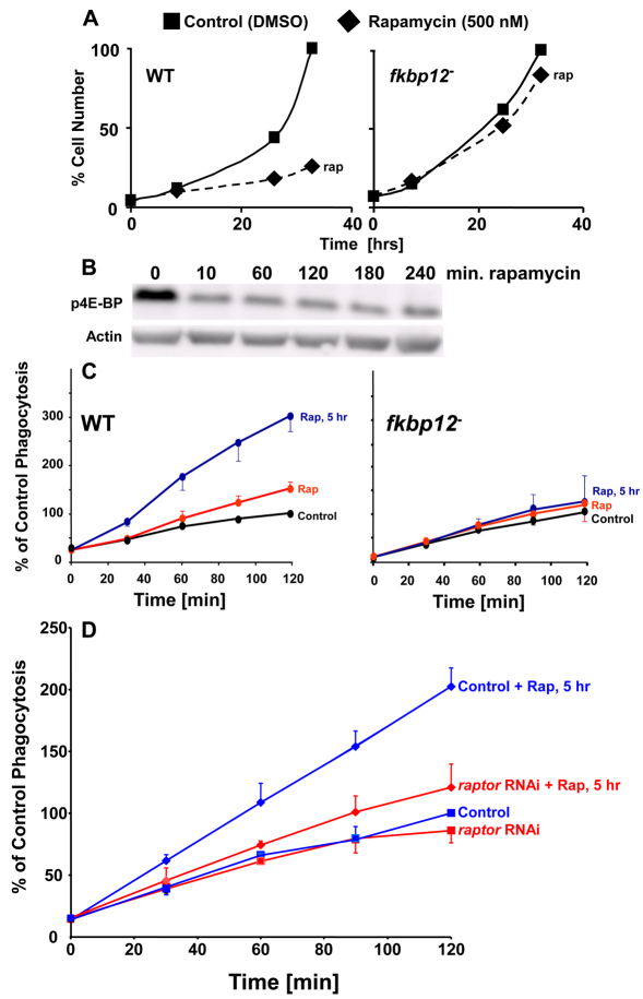
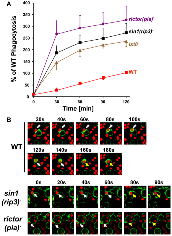
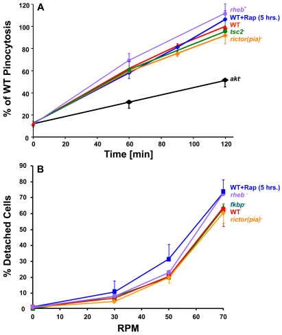
The TOR protein kinase functions in two distinct complexes, TOR complex 1 (TORC1) and 2 (TORC2). TORC1 is required for growth in response to growth factors, nutrients and the cellular energy state; TORC2 regulates AKT signaling, which can modulate cytoskeletal polarization. In its ecological niche, Dictyostelium engulf bacteria and yeast for nutrient capture. Despite the essential role of TORC1 in control of cellular growth, we show that nutrient particle capture (phagocytosis) in Dictyostelium is independent of TORC1-mediated nutrient sensing and growth regulation. However, loss of Dictyostelium TORC2 components Rictor/Pia, SIN1/RIP3 and Lst8 promotes nutrient particle uptake; inactivation of TORC2 leads to increased efficiency and speed of phagocytosis. In contrast to phagocytosis, we show that macropinocytosis, an AKT-dependent process for cellular uptake of fluid phase nutrients, is not regulated by either of the TOR complexes. The integrated and balanced regulation of TORC1 and TORC2 might be crucial in Dictyostelium to coordinate growth and energy needs with other essential TOR-regulated processes.


Figures

Fig. 1.
Fig. 1.
TORC1 activity is not correlated with phagocytosis. (A) Nutrient withdrawal induces the rapid dephosphorylation of 4E-BP. Wild-type cells were grown to log phase (<2×10⁶ cells/ml) and then transferred to starvation buffer. Whole cell protein samples were prepared at times indicated and immunoblotted using antibodies against human 4E-BP1 phosphorylated at T37 and/or T46 (p4E-BP) and actin. Experiments were repeated three times. (B,C) 4E-BP phosphorylation during phagocytosis in nutrient-rich media or in starvation buffer. Wild-type cells were grown to log phase (<2×10⁶ cells/ml). One aliquot (growing, in media) was mixed immediately with TRITC-labeled, heat-killed yeast particles, and one aliquot (starved, in buffer) was transferred to starvation buffer for 2 hours prior to receiving TRITC-labeled, heat-killed yeast particles. At the times indicated, samples were removed and monitored for (B) relative 4E-BP phosphorylation by immunoblot assay (see A) or (C) relative phagocytosis by fluorimetric analyses. Arbitrary fluorescence units were used to normalize each strain relative to the maximum obtained for wild-type in growth media within the same experiment. The data shown in B and C are from a single representative experiment. The experiment was performed three times and there was no significant difference (P=0.1) in phagocytoisis between starved and growing cells (Fig. 1C).
Fig. 2.
Fig. 2.
Raptor is required for growth, but for not phagocytosis. (A,B) Raptor is required for growth. raptor RNAi cells were grown to log phase (<2×10⁶ cells/ml) in tetracycline and diluted ~10× into fresh media with or without tetracyline as indicated. Aliquots were taken at various times to evaluate (A) relative levels of endogenous raptor mRNA (~5 kb) by RNA blot analyses (rRNAs, ribosomal RNAs used as loading control markers) and (B) relative growth rates. (C) Raptor is not required for phagocytosis. raptor RNAi cells cultured with (control) or without tetracycline (raptor RNAi) for 34–40 hours. (see A,B) were assayed for relative phagocytosis by fluorimetric analyses. Arbitrary fluorescence units were used to normalize each strain relative to the maximum obtained for the control cells (plus tetracycline) within the same experiment. Three comparisons were made; values indicate means ± s.d.; differences to each other or to wild-type or mock cells (data not shown) were not significant. For each experiment, we first confirmed growth inhibition of raptor-depleted cells after removal of tetracycline (see B). Data are from cells that also displayed suppression of Raptor expression.
Fig. 3.
Fig. 3.
Differential effects of rapamycin on growth, 4E-BP phosphorylation and phagocytosis. (A) FKBP12 is required for rapamycin to inhibit Dictyostelium growth. Wild-type (WT) and fkbp12-null cells were grown to log phase (<2×10⁶ cells/ml) and diluted ~10× into fresh media containing DMSO alone (control) or with 500 nM rapamycin. Aliquots were taken at various times to evaluate relative growth rate. (B) Rapamycin induces the rapid dephosphorylation of 4E-BP. Wild-type cells were grown to log phase (<2×10⁶ cells/ml) and rapamycin was added to 500 nM. Whole cell protein samples were taken at times indicated and immunoblotted using antibodies against human 4E-BP1 phosphorylated at T37 and/or T46 (p4E-BP) and actin. Experiments were repeated three times. (C) Long-term, but not short-term, treatment with rapamycin stimulates phagocytosis. Wild-type or fkbp12-null cells were untreated (Control) or pretreated with 500 nM rapamycin for 30 minutes (Rap) or 5 hours (Rap, 5 hr), and mixed with TRITC-labeled, heat-killed yeast particles in the continued presence (or absence) of 500 nM rapamycin. At the times indicated, samples were removed and monitored by fluorimetric analyses. Arbitrary fluorescence units were used to normalize each strain relative to the maximum obtained for untreated control cells within the same experiment. Each of the treatments was compared at least three times; values indicate means ± s.d. (see supplementary material Table S1, Fig. S1). (D) Raptor is required for long-term stimulation of phagocytosis by rapamycin. raptor RNAi cells were cultured with or without tetracycline (see Fig. 2) for 34–40 hours. and either pretreated with 500 nM rapamycin for 5 hours or not treated. The various cells were then mixed with TRITC-labeled, heat-killed yeast particles in the continued presence (or absence) of tetracycline and/or rapamycin. At the times indicated, samples were removed and monitored by fluorimetric analyses. Arbitrary fluorescence units were used to normalize each strain relative to the maximum obtained for control cells (+ tetracycline; – rapamycin) within the same experiment. Each of the treatments was compared at least three times; values indicate means ± s.d. For each experiment, we first confirmed growth inhibition of raptor-depleted cells after removal of tetracycline (see Fig. 2B). Data are from cells that also displayed suppression of Raptor expression. Statistical analysis did not reveal significant differences (P<0.05) between raptor RNAi cells and raptor RNAi cells treated with rapamycin (see supplementary material Fig. S1).
Fig. 4.
Fig. 4.
TORC2 supresses phagocytosis. (A) Wild-type (WT) and mutant strains of Dictyostelium were mixed with TRITC-labeled, heat-killed yeast particles. At the times indicated, samples were removed and monitored by fluorimetric analyses. Arbitrary fluorescence units were used to normalize each strain relative to the maximum obtained for wild-type within the same experiment. Each of the cell lines was examined at least four times; values indicate means ± s.d. (see Table 1). Differences to wild-type were statistically significant (P<0.05). (B) Actin cytoskeletal associations at the phagocytic cup do not require TORC2. The various cell lines were marked with the fluorescence F-actin binding marker GFP–ABD (green) and mixed with TRITC-labeled, heat-killed yeast particles (red). Individual cells were imaged simultaneously for GFP and rhodamine fluorescence over time using confocal microscopy. Polymerization and depolymerization of actin (green) is observed around a yeast particle (red), marked by an arrow. Although expression of GFP–ABD has a slight deleterious effect on phagocytosis, it reduces particle engulfment rates equivalently in all cell lines. Thus, even in the absence of GFP–ABD, the rictor(pia)- and sin1(rip3)-null cells exhibit proportionately more efficient phagocytosis than do wild-type controls (see Table 2). The white-to-yellow arrow transition marks the time of particle internalization, which occurs more rapidly in the sin1(rip3)- and rictor(pia)-null cells than in wild-type cells (see also Table 2).
Fig. 5.
Fig. 5.
AKT, TSC2 and Rheb differentially regulate phagocytosis. Wild-type (WT) and mutant strains of Dictyostelium were mixed with TRITC-labeled, heat-killed yeast particles. At the times indicated, samples were removed and monitored by fluorimetric analyses. Arbitrary fluorescence units were used to normalize each strain relative to the maximum obtained for wild-type within the same experiment. Each of the cell lines was examined at least three times; values indicate means ± s.d. (see Table 3); differences to wild-type for all other stains were statistically significant (P<0.05).
Fig. 6.
Fig. 6.
TORC2 and TORC1 do not regulate macropinocytosis or adhesion. (A) Wild-type (WT) and TORC2 mutant strains of Dictyostelium were mixed with TRITC–dextran at room temperature at 160 r.p.m. Wild-type cells pretreated with 500 nM rapamycin for 5 hours as indicated were also mixed with TRITC-dextran at room temperature at 160 r.p.m. At the times indicated, samples were removed and monitored by fluorimetric analyses. Arbitrary fluorescence units were used to normalize each strain relative to the maximum obtained for wild-type within the same experiment. Each of the cell lines was examined at least three times; values indicate means ± s.d. (B) Wild-type and mutant strains of Dictyostelium were allowed to adhere to tissue culture dishes as described (Khurana et al., 2005). Wild-type cells pretreated with 500 nM rapamycin for 5 hours as indicated, were also allowed to adhere to tissue culture dishes as described (Khurana et al., 2005). The dishes were then shaken for 60 minutes at varying speeds, and the percentage of unattached cells determined. Each of the cell lines was examined at least three times; values indicate means ± s.d. Differences were not statistically significant.
Fig. 7.
Fig. 7.
Activated Rheb requires TORC2, but not AKT, to suppress phagocytosis. Wild-type (WT) and mutant strains of Dictyostelium were mixed with TRITC-labeled, heat-killed yeast particles. At the times indicated, samples were removed and monitored by fluorimetric analyses. Arbitrary fluorescence units were used to normalize each strain relative to the maximum obtained for wild-type within the same experiment. Each of the cell lines was examined at least three times; values indicate means ± s.d. Differences to wild-type for all stains were statistically significant (P<0.05). (A) Expression of the GTP-bound, RhebQ64L and the non-GTP-bound RhebD60I variants have contrasting effects on phagocytosis (see Table 3). (B) Expression of the GTP-bound Rheb variant RhebQ64L suppressed phagocytosis of akt-null cells. (C) Expression of the GTP-bound Rheb variant RhebQ64L is not able to suppress phagocytosis rip3- or lst8-null cells.
Fig. 8.
Fig. 8.
Interaction of TOR pathway members for the regulation of phagocytosis. TORC2 is composed of subunits TOR, Rictor(Pia), Sin1(RIP3) and Lst8; TORC2 suppresses phagocytosis. TORC1 is composed of subunits TOR, Raptor and Lst8 and phosphorylates 4E-BP; TORC1 promotes growth. TORC1 requires Raptor for function and does not regulate phagocytosis. Rapamycin in complex with FKBP12 rapidly disrupts TORC1 stability and represses phosphorylation of 4E-BP and inhibits growth. Long-term rapamycin treatment will partially inhibit TORC2 activity and stimulate phagocytosis. This process requires FKBP12 and Raptor and cannot further stimulate phagocytosis in cells lacking Rictor(Pia) or Sin1(RIP3). In Dictyostelium, neither Rheb nor Lst8 are required for growth, but loss of either increases growth sensitivity to rapamycin (our unpublished observations); thus Rheb appears to potentiate the action of TORC1. Rheb is negatively regulated by TSC2, which is negatively regulated by the AGC kinases AKT and PKBR1. In Dictyostelium, AKT activity requires PI3K for membrane localization and coordinated phosphorylations within the kinase domain (by PDK1) and the C-terminal regulatory motif [by TORC2(PDK2)] (Liao et al., 2010) and is antagonized by PTEN. PKBR1 activity also requires phosphorylation by PDK1 and TORC2, but unlike AKT, PKBR1 does not require PI3K for membrane localization or activation (Liao et al., 2010); in Dictyostelium, PI3K and PTEN do not regulate either PDK1 or TORC2 (Liao et al., 2010). AKT and PKBR1 are basally phosphorylated at both kinase and regulatory sites in Dictyostelium growing in axenic culture (Liao et al., 2010). Although, PKBR1 requires Rictor(Pia), Sin1(RIP3) and Lst8 for both of these basal phosphorylations, AKT is basally phosphorylated regardless of Rictor(Pia), Sin1(RIP3) or Lst8 (Liao et al., 2010), perhaps suggesting a different kinase complex (PDK2?). In growing Dictyostelium, AKT is the predominant AGC kinase (Liao et al., 2010). Both AKT and PKBR1 regulate macropinocytosis, but the contribution of PKBR1 is minor. Thus, loss of TOR, Rictor(Pia), Sin1(RIP3) or Lst8, which do not effect basal AKT, has minimal effect on macropinocytosis. Rapamycin only partially inhibits AKT and PKBR1 and also has only minimal effect on macropinocytosis. Loss of AKT, PKBR1 and Rheb activates phagocytosis. Loss of TSC2 suppresses phagocytosis. Activated Rheb suppresses phagocytosis through a mechanism that is independent of AKT and PKBR1, but that requires TORC2 components Rictor(Pia), Sin1(RIP3) and Lst8. Because TORC1 activity does not regulate phagocytosis, activated Rheb could function upstream of TORC2 in this pathway.

Similar articles

Cited by

References

    1. Abraham R. T., Wiederrecht G. J. (1996). Immunopharmacology of rapamycin. Annu. Rev. Immunol. 14, 483-510 - PubMed
    1. Aspuria P. J., Sato T., Tamanoi F. (2007). The TSC/Rheb/TOR signaling pathway in fission yeast and mammalian cells: temperature sensitive and constitutive active mutants of TOR. Cell Cycle 6, 1692-1695 - PubMed
    1. Avruch J., Long X., Ortiz-Vega S., Rapley J., Papageorgiou A., Dai N. (2008). Amino Acid Regulation of TOR Complex 1. Am. J. Physiol. Endocrinol. Metab. 96, E592-E602 - PMC - PubMed
    1. Blaauw M., Linskens M. H., van Haastert P. J. (2000). Efficient control of gene expression by a tetracycline-dependent transactivator in single Dictyostelium discoideum cells. Gene 252, 71-82 - PubMed
    1. Brunn G. J., Hudson C. C., Sekulic A., Williams J. M., Hosoi H., Houghton P. J., Lawrence J. C., Jr, Abraham R. T. (1997). Phosphorylation of the translational repressor PHAS-I by the mammalian target of rapamycin. Science 277, 99-101 - PubMed

Publication types

MeSH terms

Substances

LinkOut - more resources