. 2012 Jan 1;125(Pt 1):121-32.
doi: 10.1242/jcs.087841. Epub 2012 Jan 20.

Periostin modulates myofibroblast differentiation during full-thickness cutaneous wound repair

Affiliations

Periostin modulates myofibroblast differentiation during full-thickness cutaneous wound repair

Christopher G Elliott et al. J Cell Sci. .

Abstract

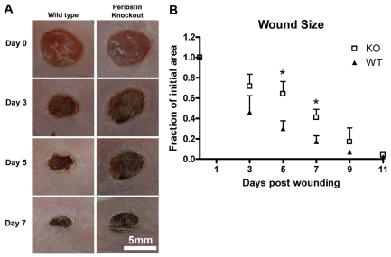
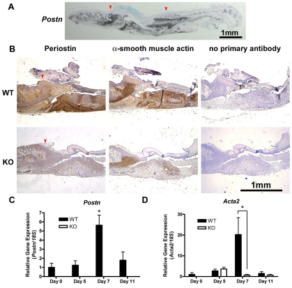
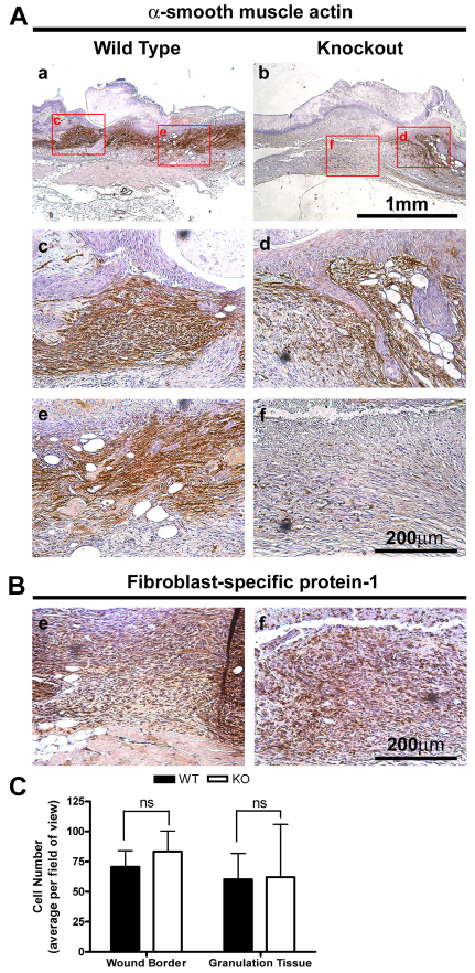
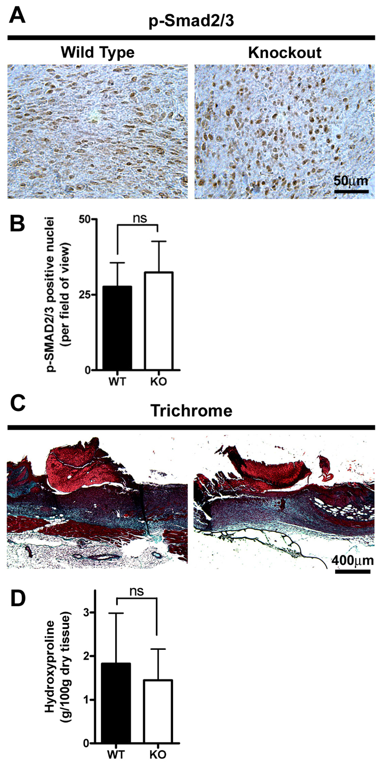
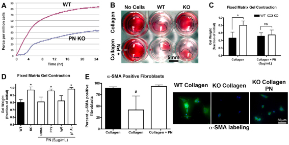
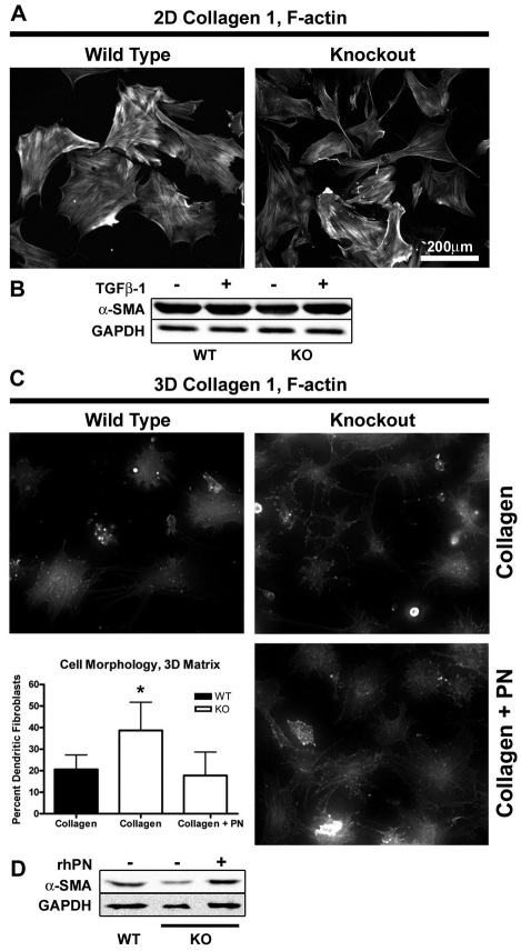
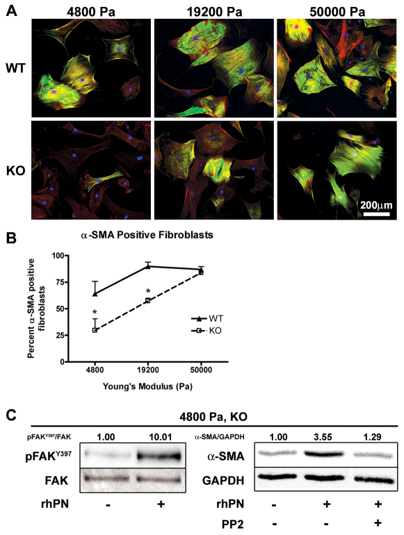
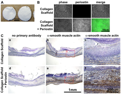
The matricellular protein periostin is expressed in the skin. Although periostin has been hypothesized to contribute to dermal homeostasis and repair, this has not been directly tested. To assess the contribution of periostin to dermal healing, 6 mm full-thickness excisional wounds were created in the skin of periostin-knockout and wild-type, sex-matched control mice. In wild-type mice, periostin was potently induced 5-7 days after wounding. In the absence of periostin, day 7 wounds showed a significant reduction in myofibroblasts, as visualized by expression of α-smooth muscle actin (α-SMA) within the granulation tissue. Delivery of recombinant human periostin by electrospun collagen scaffolds restored α-SMA expression. Isolated wild-type and knockout dermal fibroblasts did not differ in in vitro assays of adhesion or migration; however, in 3D culture, periostin-knockout fibroblasts showed a significantly reduced ability to contract a collagen matrix, and adopted a dendritic phenotype. Recombinant periostin restored the defects in cell morphology and matrix contraction displayed by periostin-deficient fibroblasts in a manner that was sensitive to a neutralizing anti-β1-integrin and to the FAK and Src inhibitor PP2. We propose that periostin promotes wound contraction by facilitating myofibroblast differentiation and contraction.


Figures

Fig. 1.
Fig. 1.
Loss of periostin results in altered wound-closure kinetics. Full-thickness excisional punch wounds were created in the skin of Postn+/+ and Postn−/− mice using a 6 mm biopsy tool. (A) Wounds were photographed at 0, 3, 5, 7 and 11 days after wounding. Four pairs of mice (four wounds per mouse) were followed for 11 days. (B) Quantification of wound area was from photographs. Postn−/− (KO) wounds are delayed in closure at days 5 and 7 (P<0.01). Data are expressed as a fraction of the initial wound area; error bars represent s.d. (*P<0.01; two-way ANOVA).
Fig. 2.
Fig. 2.
α-SMA expression is reduced in the granulation tissue of Postn−/− mice. (A) Postn message detection in day 7 Postn+/+ wounds by in situ hybridization, showing periostin expression selectively in the wound. Arrowheads indicate the wound borders. (B) Histological analysis of day 7 wounds from Postn+/+ (WT) and Postn−/− (KO) animals. Sections were incubated with antibodies against periostin or α-SMA. Detection was with peroxidase-conjugated secondary antibodies and DAB. Day 7 Postn−/− wounds have reduced α-SMA staining (n=5). (C) Healthy skin or wound tissue biopsies were analyzed for Postn and (D) Acta2 mRNA by RT-qPCR. Target gene expression was normalized to 18S using the ΔΔCt method. Postn expression is significantly increased at day 7 (P<0.001, n=5). Postn mRNA was not detected in Postn−/− specimens. Increased Acta2 expression was observed at day 7 in Postn+/+ wounds, but not in Postn−/− wounds (P<0.001, n=5). Data are expressed relative to day 0 Postn+/+ expression; error bars represent s.e.m. (*P<0.001; two-way ANOVA).
Fig. 3.
Fig. 3.
Decreased α-SMA expression is restricted to the granulation tissue of Postn−/− mice. (A) Histological analysis of day 7 wounds from Postn+/+ and Postn−/− animals. Sections were incubated with an antibody for α-SMA (B) or fibroblast-specific protein-1. Detection was with peroxidase conjugated secondary antibodies and DAB. α-SMA is absent in the granulation tissue of Postn−/− wounds, but is present at the wound border (n=4). Granulation tissue is dominated by fibroblasts in both Postn+/+ and Postn−/− day 7 wounds. (C) Quantification of cell number at the wound border and within the granulation tissue in high magnification fields of view. Cell number was not different between Postn+/+ (WT) and Postn−/− (KO) wounds at either location (P=0.28). Data is expressed as mean number of cells per field of view; error bars represent s.d. (two-way ANOVA).
Fig. 4.
Fig. 4.
Canonical TGFβ signaling is not altered in Postn−/− fibroblasts. (A) Histological analysis of day 7 wounds from Postn+/+ and Postn−/− animals. Sections were incubated with an antibody for phosphorylated Smad2/3 (p-Smad2/3). Detection was with peroxidase conjugated secondary antibodies and DAB (n=3). (B) Numbers of positively stained nuclei per high power field of view were not significantly different between Postn+/+ (WT) and Postn−/− (KO) wounds (P=0.37; Student's t-test). (C) Masson's trichrome staining of day 7 wound sections. (D) Hydroxyproline content (g/100 g dry tissue) of excised day 7 wounds was not different between Postn+/+ and Postn−/− animals (P=0.60; Student's t-test). Data are expressed as means; error bars represent s.d.
Fig. 5.
Fig. 5.
Exogenous periostin is sufficient to induce a contractile phenotype. (A) The effect of periostin (PN) deletion on the ability of dermal fibroblasts to exert contractile force in a fixed, tethered floating collagen gel lattice was investigated using a culture force monitor. Forces generated by fibroblasts were measured over 24 hours; a representative trace is shown (n=3). (B) Cells contracted collagen gels over an additional 24 hours at 37°C, 5% CO2. (C) Gel contraction was quantified by loss of gel weight, compared with gels lacking cells. Postn−/− (KO) fibroblasts were unable to significantly contract collagen gels. Note that Postn+/+ (WT) fibroblasts were able to contract collagen gels. Addition of 5 μg/ml rhPN to the collagen gels rescued the contractile ability of Postn−/− fibroblasts (n=3). Data is expressed as a fraction of the initial gel weight; error bars represent s.d. (*P<0.05; two-way ANOVA). (D) Gels were treated with 10 μM PP2 (or DMSO vehicle) or 10 μg/ml β1-integrin blocking antibody (mouse IgG for controls). Data are expressed as a fraction of the initial gel weight; error bars represent s.d. (*P<0.05; one-way ANOVA, n=3). A Dunnett's multiple comparison test was used where Postn−/−, rhPN and DMSO were used as the reference group. Extracellular periostin influences contractility through a β1-integrin- and FAK-dependent mechanism in vitro. (E) Fluorescent labeling of fibroblast populated collagen gels for α-SMA (green) and nuclei (blue). α-SMA-positive cells were counted in high magnification fields of view. Percentage of α-SMA-positive cells was significantly reduced in Postn−/− fibroblast populated gels (P<0.01; n=3). Addition of 5 μg/ml rhPN to the collagen gels restored the percentage of α-SMA-positive cells. Data are expressed as mean; error bars represent s.d. (#P<0.01; one-way ANOVA).
Fig. 6.
Fig. 6.
Periostin influences fibroblast morphology in 3D culture but not 2D culture. Isolated dermal fibroblasts were seeded on collagen-coated tissue culture plates or onto precast anchored collagen gels. Cells were incubated for 48 hours before fixation or harvesting lysates. (A) Filamentous actin was visualized with Rhodamine-conjugated Phalloidin. Distinct stress fibers were observed in both Postn+/+ and Postn−/− fibroblasts (n=3). (B) Western blot analysis of lysates was carried out to quantify the level of α-SMA. Equal loading was confirmed by blotting for GAPDH. No difference was detected between Postn+/+ (WT) and Postn−/− (KO) fibroblasts. (C) Fibroblasts seeded on 3D collagen gels were labeled with Rhodamine-conjugated phalloidin and assessed for cell morphology (n=3). Postn−/− fibroblasts were more likely to adopt a dendritic phenotype, characterized by a lack of stress fibers and extension of thin branching cytoplasmic extensions (P<0.01). Addition of 5 μg/ml rhPN to the gels restored the percentage of dendritic fibroblasts to Postn+/+ levels. Data is expressed as mean; error bars represent s.d. (*P<0.01; one-way ANOVA). (D) 3D collagen gels were homogenized and cells were lysed by sonication. α-SMA levels were assessed by western blot.
Fig. 7.
Fig. 7.
Periostin facilitates α-SMA expression on a soft substrate, but is compensated for by increased substrate stiffness. Isolated dermal fibroblasts were seeded on collagen-coated flexible polyacrylamide substrates with Young's moduli of 4800, 19,200 or 50,000 Pa. Cells were incubated for 48 hours before fixation. (A) Fibroblasts were fluorescently labeled for α-SMA (green), filamentous actin (red) and nuclei (blue). α-SMA positive cells were counted from high magnification fields of view. (B) Percentage of α-SMA-positive cells was significantly reduced in Postn−/− (KO) fibroblasts grown on the soft 4800 and 19,200 Pa substrates (P<0.05, n=3). On the stiff 50,000 Pa substrates, however, the proportion of α-SMA-positive Postn−/− fibroblasts was equivalent to that of Postn+/+ (WT) fibroblasts. Data are expressed as means; error bars represent s.d. (*P<0.05; two-way ANOVA). (C) Western blot analysis of lysates from Postn−/− fibroblasts grown on 4800 Pa substrates was carried out to quantify the level of FAK-P [at Y397; pFAK(Y397)] and α-SMA. Loading was corrected by blotting for total FAK and GAPDH. pFAKY397/FAK and α-SMA/GAPDH indicate results of densitometry, relative to control. Incorporation of rhPN on 4800 Pa substrates resulted in an increase in phosphorylated FAK(Y397) and α-SMA protein. Increased α-SMA was attenuated by 10 μM PP2.
Fig. 8.
Fig. 8.
Delivery of recombinant periostin via electrospun collagen scaffolds recovers α-SMA expression in Postn−/− mice. (A) Collagen type 1 electrospun scaffolds, with and without rhPN, were cut into 6 mm disks to match the size of punch wounds. (B) Periostin (green) protein was detected throughout the collagen + periostin scaffolds, but not the control collagen scaffolds. (C) Punch wounds were created in Postn−/− animals and scaffolds were immediately laid into the wounds. Half of the wounds received two control (collagen only) scaffolds and the other half of the wounds received two collagen and periostin scaffolds. Wounds were harvested for IHC at day 7. Addition of periostin to Postn−/− wounds by electrospun collagen scaffolds resulted in a marked increase in α-SMA immunoreactivity throughout the granulation tissue, when compared with that in wounds receiving control collagen scaffolds (n=3).

References

    1. Aplin J. D., Hughes R. C. (1981). Protein-derivatised glass coverslips for the study of cell-to substratum adhesion. Anal. Biochem. 113, 144-148 - PubMed
    1. Arora P. D., Narani N., McCulloch C. A. (1999). The compliance of collagen gels regulates transforming growth factor-beta induction of alpha-smooth muscle actin in fibroblasts. Am. J. Pathol. 154, 871-882 - PMC - PubMed
    1. Babu M., Diegelmann R., Oliver N. (1992). Keloid fibroblasts exhibit an altered response to TGF-beta. J. Invest. Dermatol. 99, 650-655 - PubMed
    1. Bao S., Ouyang G., Bai X., Huang Z., Ma C., Liu M., Shao R., Anderson R. M., Rich J. N., Wang X. F. (2004). Periostin potently promotes metastatic growth of colon cancer by augmenting cell survival via the Akt/PKB pathway. Cancer Cell 5, 329-339 - PubMed
    1. Baril P., Gangeswaran R., Mahon P. C., Caulee K., Kocher H. M., Harada T., Zhu M., Kalthoff H., Crnogorac-Jurcevic T., Lemoine N. R. (2007). Periostin promotes invasiveness and resistance of pancreatic cancer cells to hypoxia-induced cell death: role of the beta4 integrin and the PI3k pathway. Oncogene 26, 2082-2094 - PubMed

Publication types

MeSH terms