. 2012 May;37(5):511-7.
doi: 10.1097/SHK.0b013e318249cfa2.

Splenocyte apoptosis and autophagy is mediated by interferon regulatory factor 1 during murine endotoxemia

Affiliations

Splenocyte apoptosis and autophagy is mediated by interferon regulatory factor 1 during murine endotoxemia

Lemeng Zhang et al. Shock. 2012 May.

Abstract

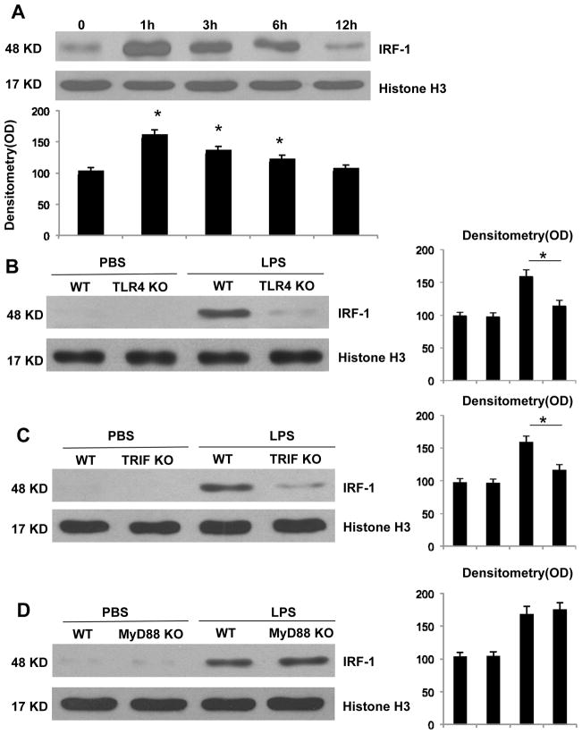
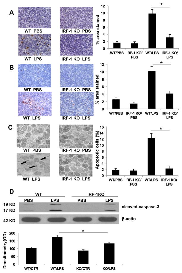
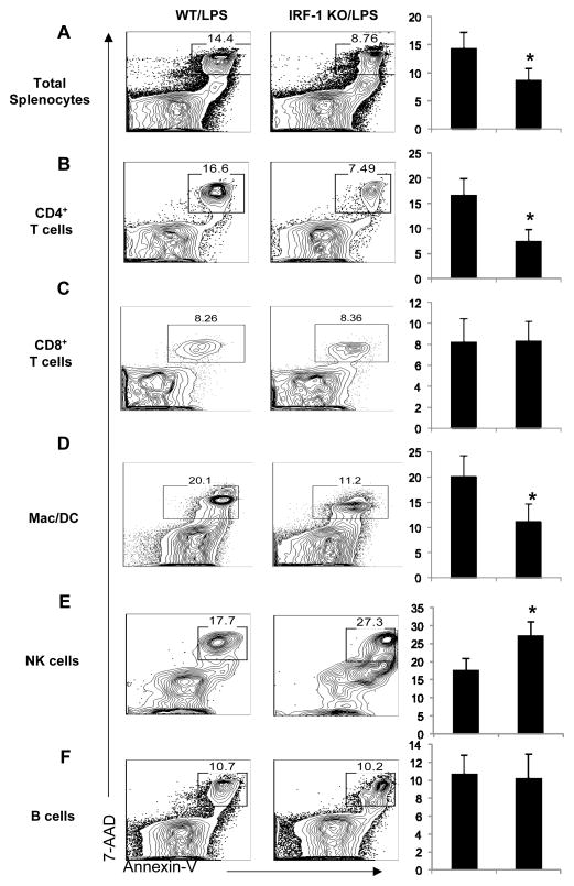
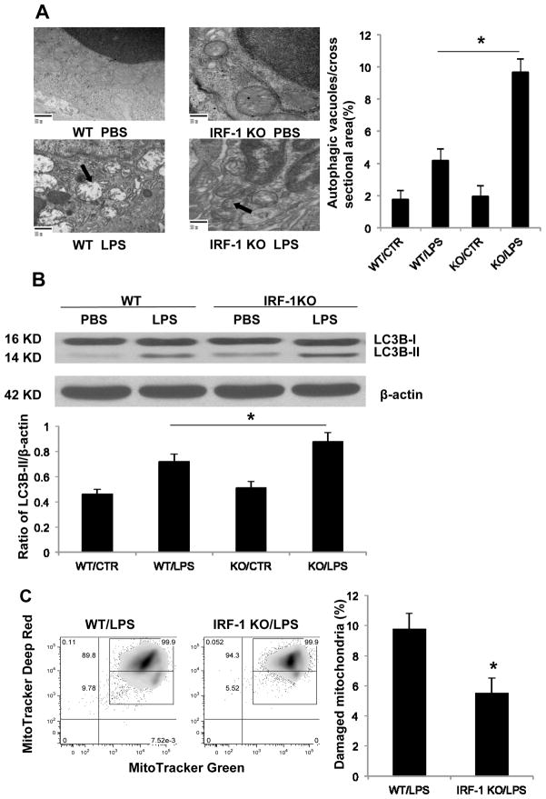
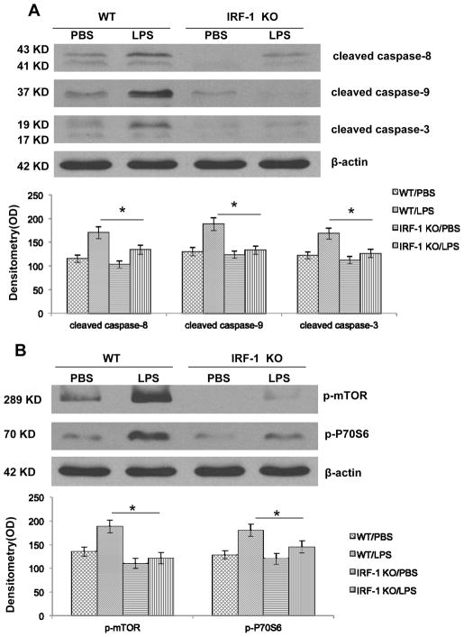
Sepsis-induced lymphocyte and dendritic cell apoptosis contributes to immunosuppression, which results in an inability to eradicate the primary infection as well as a propensity to acquire new, secondary infections. Another cellular process, autophagy, is also activated in immune cells and plays a protective role. In the present study, we demonstrate that interferon regulatory factor 1 (IRF-1) regulates both immune cell apoptosis and autophagy in a murine endotoxemia model. Interferon regulatory factor 1 is activated at an early phase through a Toll-like receptor 4-dependent, myeloid differentiation primary response gene 88-independent manner in splenocytes. Furthermore, IRF-1 knockout (KO) mice are protected from a lethal endotoxemia model. This protection is associated with decreased apoptosis and increased autophagy in splenocytes. Interferon regulatory factor 1 KO mice experience decreased apoptotic cell loss, especially in CD4⁺ T lymphocytes and myeloid antigen-presenting cells. Meanwhile, IRF-1 KO mice demonstrate increased autophagy and improved mitochondrial integrity. This increased autophagy in KO mice is attributable, at least in part, to deactivation of mammalian target of rapamycin/P70S6 signaling--a main negative regulator of autophagy. Therefore, we propose a novel role for IRF-1 in regulating both apoptosis and autophagy in splenocytes in the setting of endotoxemia with IRF-1 promoting apoptosis and inhibiting autophagy.


Figures

Figure 1
Figure 1. IRF-1 activation at early phase and is mediated through the TLR4-TRIF dependent manner in response to LPS stimulation
(A) After LPS injection at predetermined time points, splenocytes were isolated from WT mice. Nuclear proteins were isolated and IRF-1 activation was analyzed by Western blot. WT and TLR4 KO (B), TRIF KO (C), MyD88 KO (D) mice were injected with PBS or LPS for 1 h and splenocytes were isolated. Nuclear IRF-1 was analyzed by Western blot. *, P<0.05; results are representative of 3 separate independent experiments.
Figure 2
Figure 2. IRF-1 KO mice have decreased apoptosis in splenocytes following LPS exposure
Permanent blocks of splenic tissue obtained from WT and IRF-1 KO mice 16 h following PBS or LPS (20 mg/kg) injection were sectioned and (A) TUNEL staining or (B) cleaved caspase-3 immunohistochemistry staining was performed (magnitude X 200). Positive staining was presented as percentage of stained area over total area (% area stained). (C) Splenic tissue obtained from WT and IRF-1 KO mice 16 h following PBS or LPS (20 mg/kg) injection was imaged by transmission electron microscope (magnitude X 5000). Arrow points to apoptotic bodies. Percentage of apoptotic cells among total cells was used for apoptosis quantification. (D) WT and IRF-1 KO mice were injected with PBS or LPS (20 mg/kg) for 16 h and splenocytes were isolated. Caspase-3 cleavage was analyzed by Western blot. *, P<0.05; results are representative of 3 separate independent experiments.
Figure 3
Figure 3. Apoptosis is selectively reduced in splenic CD4+ T lymphocytes and myeloid APC in IRF-1 KO mice following LPS exposure
WT and IRF1-KO mice were challenged with LPS (20 mg/kg) as described and total splenocytes were harvested. Apoptotic cells (Annexin V+7-AAD+) were detected by flow cytometry. (A) Total splenocytes, (B) CD3+CD4+ T cells, (C) CD3+CD8+ T cells, (D) CD11b+NK1.1- myeloid APC including dendritic cells and macrophages, (E) CD11b+NK1.1+ NK cells, and (F) B220+ B cells were analyzed. The percent of apoptotic cells was quantified for each cell population. *, P<0.05; results are representative of 3 separate independent experiments.
Figure 4
Figure 4. Splenocytes from IRF-1 KO mice demonstrate increased autophagy compared to WT mice following LPS exposure
(A) Splenic tissue obtained from WT and IRF-1 KO mice 16 h following PBS or LPS (20 mg/kg) injection imaged by transmission electron microscope (magnitude X 20000). Arrows point to damaged mitochondria and autophagy. Determining the number of autophagic vacuoles per cross-sectioned cell was used for autophagy quantification. (B) WT and IRF-1 KO mice were injected with PBS or LPS (20 mg/kg) for 16 h and splenocytes were isolated. LC3B-II was analyzed by Western blot. (C) WT and IRF-1 KO mice were injected with LPS (20 mg/kg) for 16 h and splenocytes were isolated and stained with 200 nM MitoTracker Deep Red and MitoTracker Green for 45 min at 37°C. Representative flow cytometry plots are represented. *, P<0.05; results are representative of 3 separate independent experiments.
Figure 5
Figure 5. IRF-1 activation promotes both intrinsic and extrinsic apoptotic pathway and inhibits autophagy by modulating mTOR/P70S6 activity
(A) WT and IRF-1 KO mice were injected with PBS or LPS (20 mg/kg) for 16 h and splenocytes were isolated. Caspase-3, -8, -9 cleavage was analyzed by Western blot. (B) WT and IRF-1 KO mice were injected with PBS or LPS (20 mg/kg) for 16 h and splenocytes were isolated. mTOR and P70S6 activation were analyzed by Western blot. *, P<0.05; results are representative of 3 separate independent experiments.

Similar articles

Cited by

References

    1. Aneja R, Fink MP. Promising therapeutic agents for sepsis. Trends Microbiol. 2007;15(1):31–7. - PubMed
    1. Park BS, Song DH, Kim HM, Choi BS, Lee H, Lee JO. The structural basis of lipopolysaccharide recognition by the TLR4-MD-2 complex. Nature. 2009;458(7242):1191–5. - PubMed
    1. Chopra M, Sharma AC. Distinct cardiodynamic and molecular characteristics during early and late stages of sepsis-induced myocardial dysfunction. Life Sci. 2007;81(4):306–16. - PMC - PubMed
    1. Xiao H, Siddiqui J, Remick DG. Mechanisms of mortality in early and late sepsis. Infect Immun. 2006;74(9):5227–35. - PMC - PubMed
    1. Hotchkiss RS, Swanson PE, Freeman BD, Tinsley KW, Cobb JP, Matuschak GM, Buchman TG, Karl IE. Apoptotic cell death in patients with sepsis, shock, and multiple organ dysfunction. Crit Care Med. 1999;27(7):1230–51. - PubMed

Publication types

MeSH terms

Substances