Recapitulation of the Achilles tendon mechanical properties during neonatal development: a study of differential healing during two stages of development in a mouse model
- PMID: 22267191
- PMCID: PMC3265027
- DOI: 10.1002/jor.21542
Recapitulation of the Achilles tendon mechanical properties during neonatal development: a study of differential healing during two stages of development in a mouse model
Abstract
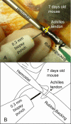
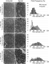
During neonatal development, tendons undergo a well-orchestrated process whereby extensive structural and compositional changes occur in synchrony to produce a normal tissue. Conversely, during the repair response to injury, structural and compositional changes occur, but a mechanically inferior tendon is produced. As a result, developmental processes have been postulated as a potential paradigm through which improved adult tissue healing may occur. By examining injury at distinctly different stages of development, vital information can be obtained into the structure-function relationships in tendon. The mouse is an intriguing developmental model due to the availability of assays and genetically altered animals. However, it has not previously been used for mechanical analysis of healing tendon due to the small size and fragile nature of neonatal tendons. The objective of this study was to evaluate the differential healing response in tendon at two distinct stages of development through mechanical, compositional, and structural properties. To accomplish this, a new in vivo surgical model and mechanical analysis method for the neonatal mouse Achilles tendons were developed. We demonstrated that injury during early development has an accelerated healing response when compared to injury during late development. This accelerated healing model can be used in future mechanistic studies to elucidate the method for improved adult tendon healing.
Copyright © 2011 Orthopaedic Research Society.
Figures


Similar articles
-
Mechanical property changes during neonatal development and healing using a multiple regression model.J Biomech. 2012 Apr 30;45(7):1288-92. doi: 10.1016/j.jbiomech.2012.01.030. Epub 2012 Feb 28. J Biomech. 2012. PMID: 22381737 Free PMC article.
-
Mechanical, compositional, and structural properties of the post-natal mouse Achilles tendon.Ann Biomed Eng. 2011 Jul;39(7):1904-13. doi: 10.1007/s10439-011-0299-0. Epub 2011 Mar 23. Ann Biomed Eng. 2011. PMID: 21431455 Free PMC article.
-
The injury response of aged tendons in the absence of biglycan and decorin.Matrix Biol. 2014 Apr;35:232-8. doi: 10.1016/j.matbio.2013.10.008. Epub 2013 Oct 21. Matrix Biol. 2014. PMID: 24157578 Free PMC article.
-
Tendon healing: repair and regeneration.Annu Rev Biomed Eng. 2012;14:47-71. doi: 10.1146/annurev-bioeng-071811-150122. Annu Rev Biomed Eng. 2012. PMID: 22809137 Review.
-
Small Leucine-Rich Proteoglycans in Tendon Wound Healing.Adv Wound Care (New Rochelle). 2022 Apr;11(4):202-214. doi: 10.1089/wound.2021.0069. Epub 2021 Dec 31. Adv Wound Care (New Rochelle). 2022. PMID: 34978952 Review.
Cited by
-
Preclinical tendon and ligament models: Beyond the 3Rs (replacement, reduction, and refinement) to 5W1H (why, who, what, where, when, how).J Orthop Res. 2023 Oct;41(10):2133-2162. doi: 10.1002/jor.25678. Epub 2023 Aug 28. J Orthop Res. 2023. PMID: 37573480 Free PMC article. Review.
-
Large Animal Models in Regenerative Medicine and Tissue Engineering: To Do or Not to Do.Front Bioeng Biotechnol. 2020 Aug 13;8:972. doi: 10.3389/fbioe.2020.00972. eCollection 2020. Front Bioeng Biotechnol. 2020. PMID: 32903631 Free PMC article. Review.
-
Tendon Cell Regeneration Is Mediated by Attachment Site-Resident Progenitors and BMP Signaling.Curr Biol. 2020 Sep 7;30(17):3277-3292.e5. doi: 10.1016/j.cub.2020.06.016. Epub 2020 Jul 9. Curr Biol. 2020. PMID: 32649909 Free PMC article.
-
Improved biomechanical and biological outcomes in the MRL/MpJ murine strain following a full-length patellar tendon injury.J Orthop Res. 2015 Nov;33(11):1693-703. doi: 10.1002/jor.22928. Epub 2015 May 25. J Orthop Res. 2015. PMID: 25982892 Free PMC article.
-
Temporal application of lysyl oxidase during hierarchical collagen fiber formation differentially effects tissue mechanics.Acta Biomater. 2023 Apr 1;160:98-111. doi: 10.1016/j.actbio.2023.02.024. Epub 2023 Feb 21. Acta Biomater. 2023. PMID: 36822485 Free PMC article.
References
-
- Gelberman RH, Vandeberg JS, Manske PR, Akeson WH. The early stages of flexor tendon healing: a morphologic study of the first fourteen days. J Hand Surg [Am] 1985;10(6 Pt 1):776–84. - PubMed
-
- Butler DL, Juncosa N, Dressler MR. Functional efficacy of tendon repair processes. Annu Rev Biomed Eng. 2004;6:303–29. - PubMed
-
- Favata M, Beredjiklian PK, Zgonis MH, et al. Regenerative properties of fetal sheep tendon are not adversely affected by transplantation into an adult environment. J Orthop Res. 2006;24(11):2124–32. - PubMed
-
- Parry DA, Barnes GR, Craig AS. A comparison of the size distribution of collagen fibrils in connective tissues as a function of age and a possible relation between fibril size distribution and mechanical properties. Proc R Soc Lond B Biol Sci. 1978;203(1152):305–21. - PubMed
Publication types
MeSH terms
Substances
Grants and funding
LinkOut - more resources
Full Text Sources
Medical
Research Materials

