Review
. 2013 Feb;20(2):140-53.
doi: 10.1177/1933719111424454. Epub 2012 Jan 19.

Extracellular matrix dynamics and fetal membrane rupture

Affiliations
Review

Extracellular matrix dynamics and fetal membrane rupture

Jerome F Strauss 3rd. Reprod Sci. 2013 Feb.

Abstract

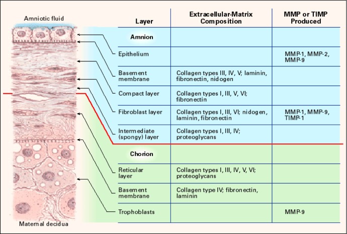
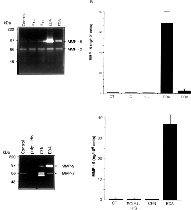
The extracellular matrix (ECM) plays an important role in determining cell and organ function: (1) it is an organizing substrate that provides tissue tensile strength; (2) it anchors cells and influences cell morphology and function via interaction with cell surface receptors; and (3) it is a reservoir for growth factors. Alterations in the content and the composition of the ECM determine its physical and biological properties, including strength and susceptibility to degradation. The ECM components themselves also harbor cryptic matrikines, which when exposed by conformational change or proteolysis have potent effects on cell function, including stimulating the production of cytokines and matrix metalloproteinases (MMPs). Collectively, these properties of the ECM reflect a dynamic tissue component that influences both tissue form and function. This review illustrates how defects in ECM synthesis and metabolism and the physiological process of ECM turnover contribute to changes in the fetal membranes that precede normal parturition and contribute to the pathological events leading to preterm premature rupture of membranes (PPROM).


Conflict of interest statement

Declaration of Conflicting Interests: The author declared no potential conflicts of interest with respect to the research, authorship, and/or publication of this article.

Figures

Figure 1.
Figure 1.
Structure of the human fetal membranes and localization of ECM components and matrix degrading enzymes (MMP) and inhibitors (TIMP). ECM indicates extracellular matrix; MMP, matrix metalloproteinase; TIMP, tissue inhibitor of metalloproteinase. Reprinted from Parry and Strauss.
Figure 2.
Figure 2.
Zymogram demonstrating amniotic fluid levels of gelatinases MMP-2 (72 kDa) and MMP-9 (97 kDa) from a pregnant baboon. Sampling dates are noted above each lane. The female delivered on 9/28. MMP indicates matrix metalloproteinase.
Figure 3.
Figure 3.
Zymogram demonstrating regional expression of gelatinases, MMP-2 (72 kDa), and MMP-9 (97 kDa) in the portion of the human amnion lying over the cervix in labored membranes (top panel) and the relative lack of regional induction in membranes collected from a nonlaboring participant (bottom panel). A indicates amnion; C, chorion; P, periplacental; M, mid-region; R, putative rupture site; MMP, matrix metalloproteinase.
Figure 4.
Figure 4.
Schematic of the domain structure of fibronectin splice variants (upper panel) showing oncofetal fibronectin exons in yellow, and the presence of EDA-containing fibronectin fragments detected by Western blotting using monoclonal antibody IST-9 (recognizing EDA) in cervical vaginal fluid from women in preterm labor (lower panel A), and neutralization of the antibody with recombinant EDA (lower panel B). Note that oncofetal fibronectin monomers have a molecular weight of 250 000 and are not shown in the blots. EDA indicates extra domain A.
Figure 5.
Figure 5.
Induction of gelatinase expression in cultured THP-1 cells exposed to the indicated recombinant His-tagged fibronectin domains. The THP-1 cells (5 × 10⁵ cells/well) were either untreated (control) or treated with 1 μM of different recombinant fibronectin type III repeats, poly-l-histidine, or cellular fibronectin (CFN) for 48 hours. A, Gelatinase activities released from THP-1 cells analyzed using gelatin zymography. B, MMP-9 in conditioned medium measured by ELISA and expressed as the mean ± SD from triplicate cultures. MMP indicates matrix metalloproteinase; ELISA, enzyme-linked immunosorbent assay; SD, standard deviation. Reprinted from Okamura et al. 5

Similar articles

Cited by

References

    1. Frantz C, Stewart KM, Weaver VM. The extracellular matrix at a glance. J Cell Sci. 2010;123(Pt 24):4195–4200 - PMC - PubMed
    1. Egeblad M, Rasch MG, Weaver VM. Dynamic interplay between the collagen scaffold and tumor evolution. Curr Opin Cell Biol. 2010;22(5):697–706 - PMC - PubMed
    1. Hadler-Olsen E, Fadnes B, Sylte I, Uhlin-Hansen L, Winberg JO. Regulation of matrix metalloproteinase activity in health and disease. FaEBS J. 2011;278(1):28–45 - PubMed
    1. Whitley GS, Cartwright JE. Cellular and molecular regulation of spiral artery remodelling: lessons from the cardiovascular field. Placenta. 2010;31(6):465–474 - PMC - PubMed
    1. Parry S, Strauss JF., 3rd Premature rupture of the fetal membranes. N Engl J Med. 1998;338(10):663–670 - PubMed

Publication types

MeSH terms

Substances

LinkOut - more resources