. 2012 Mar 23;287(13):10210-10222.
doi: 10.1074/jbc.M111.327809. Epub 2012 Jan 20.

Structure of complement C6 suggests a mechanism for initiation and unidirectional, sequential assembly of membrane attack complex (MAC)

Affiliations

Structure of complement C6 suggests a mechanism for initiation and unidirectional, sequential assembly of membrane attack complex (MAC)

Alexander E Aleshin et al. J Biol Chem. .

Abstract

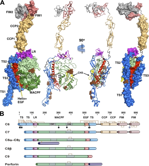
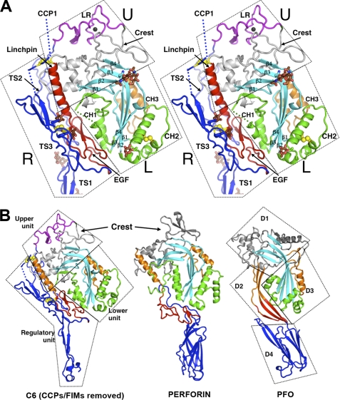
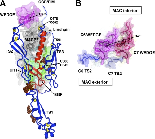
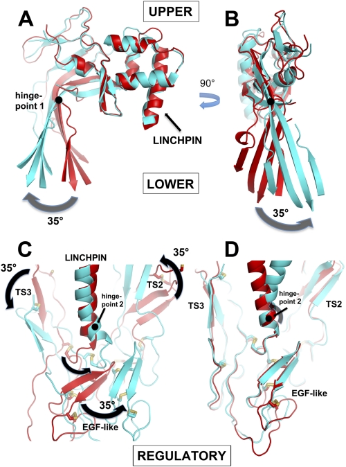
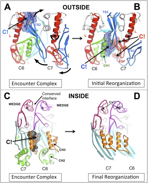
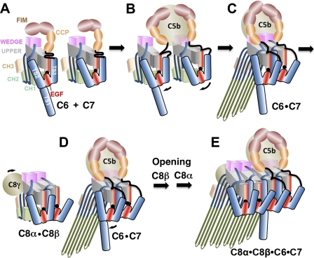
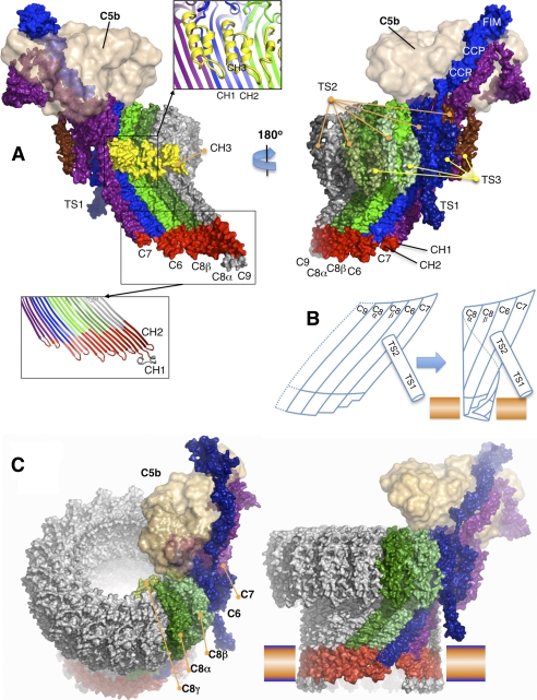
The complement membrane attack complex (MAC) is formed by the sequential assembly of C5b with four homologous proteins as follows: one copy each of C6, C7, and C8 and 12-14 copies of C9. Together these form a lytic pore in bacterial membranes. C6 through C9 comprise a MAC-perforin domain flanked by 4-9 "auxiliary" domains. Here, we report the crystal structure of C6, the first and longest of the pore proteins to be recruited by C5b. Comparisons with the structures of the C8αβγ heterodimer and perforin show that the central domain of C6 adopts a "closed" (perforin-like) state that is distinct from the "open" conformations in C8. We further show that C6, C8α, and C8β contain three homologous subdomains ("upper," "lower," and "regulatory") related by rotations about two hinge points. In C6, the regulatory segment includes four auxiliary domains that stabilize the closed conformation, inhibiting release of membrane-inserting elements. In C8β, rotation of the regulatory segment is linked to an opening of the central β-sheet of its clockwise partner, C8α. Based on these observations, we propose a model for initiation and unidirectional propagation of the MAC in which the auxiliary domains play key roles: in the assembly of the C5b-8 initiation complex; in driving and regulating the opening of the β-sheet of the MAC-performin domain of each new recruit as it adds to the growing pore; and in stabilizing the final pore. Our model of the assembled pore resembles those of the cholesterol-dependent cytolysins but is distinct from that recently proposed for perforin.


Figures

FIGURE 1.
FIGURE 1.
Crystal structure and domain organization of C6. A, surface and secondary structure presentations of C6 (orthogonal orientations). The disordered residues between modules TS3 and CCP1, CCP2 and FIM1, and the entire FIM2 are colored gray-black; the sugars of the glycosylation sites are shown as brown spheres. B, schematic presentation of the primary structural features of the complement MAC components and perforin. The disulfide bonds and the glycosylation sites of C6 are shown as brown brackets and black hexagons. Perforin contains a distinct membrane-binding C-terminal domain.
FIGURE 2.
FIGURE 2.
Structure of C6 core and its interaction with auxiliary domains. A, stereo view of the core fragment of C6 presented as a secondary structure ribbon. The rigid-body units are enclosed with boxes and labeled U (upper), L (lower), and R (regulatory). The regulatory unit consists of EGF (red), TS1, TS2, and TS3 modules (blue). The upper unit contains the LR module (magenta) and the upper fragment of MACPF, including the linchpin helix (red). The lower unit contains the lower fragment of MACPF including CH1-CH2 (green) and CH3 (orange). Glycosylation sites are shown as brown sticks. Two disulfide bonds linking TS3 to MACPF and EGF are shown as yellow balls. B, comparison of C6 (lacking CCPs and FIMs) with perforin (PDB code 2NSJ) and a member of the CDC family, PFO (PDB code 1PFO). The domains of PFO are designated D1 to D4. D1 and D3 are analogous to the upper and lower domains of C6. The linchpin helices (in orange) and the EGF domains (in red) of C6 and perforin have some functional analogy with domain D2 of PFO, but PFO and perforin lack the regulatory functions provided by the auxiliary domains of C6. D4 may be identified with TS1 of C6 on structural and possibly functional grounds. β-Sheets are in cyan; CH1 and CH2 are in green; CH3 is orange, and the rest of the domain is gray. TS1-TS3 of C6 and the membrane-binding domains of perforin and PFO are in blue.
FIGURE 3.
FIGURE 3.
Interactions between the Y-shaped regulatory module and MACPF and a model for packing of two wedge domains. A, regulatory module is colored in blue (TS1–3) and orange (EGF domains). The MACPF core is covered with a semi-transparent surface, except for the linchpin helix. The LR domain and Crest domain of MACPF, which collectively form a wedge-shaped unit, are covered with a magenta surface. The mannose rings of C1-glycosylated Trp-547 and Trp-550 and the O-glycosylation site (glucose-fucose) of Thr-17 are shown as brown balls. Selected intra- and interdomain (numbered) disulfide bonds are shown as yellow balls. Hydrophobic side chains of TS2 and TS3 that contact the EGF domain are labeled. The TS3-CCP1 linker (residues 591–620, shown as dashed lines) is disordered everywhere except for three amino acids around the Cys-478–Cys-602 disulfide bond. The second disulfide bond Cys-500–Cys-549 attaches TS3 to EGF. CCP modules and FIMs are omitted. B, model of two “wedge” domains (C6 and C7, viewed from the “top”), forming two putative building blocks that create the upper surface of the growing pore. Note the shape complementarity and curvature, which places the TS2 modules on the MAC exterior. Ca2+ ions are labeled. The view includes only the tops of the subunits sliced near to the ends of the TS2 modules.
FIGURE 4.
FIGURE 4.
Comparison of C6 with C8α and C8β illustrating the existence of three rigid-body units connected by two hinge points. A and B, two orthogonal views (“side” and “inside” views) of MACPFs of C6 (red) and C8α (cyan) after superposition of their UPPER segments. The arrows indicate the large rigid-body rotation (by ∼35°) of the LOWER segment, about an axis (hinge point 1) shown as a black circle, represented by the lower half of the β-sheet (i.e. the CH1-CH3 clusters follow this movement but have been omitted for clarity; see also supplemental Fig. 7). Note that the sheet both “opens” (A) and “twists” (B). When C6 is compared with C8β (data not shown), there is a small opening of the sheet, but it does not twist. C, comparison of C6 (red) and C8β (cyan) after superposition of their upper segments, illustrating the ∼35° rotation of the REGULATORY segment (TS2+EGF+TS3) around hinge point 2, at the end of the linchpin helix. The view is the same as in B but is from the outside of the predicted pore. Note how the motion of the REGULATORY domain correlates with the twist of the β-sheet in B. When C6 is compared with C8α (data not shown), the motions are similar but smaller in magnitude. D, same view as in C, but here the overlay is on the REGULATORY domains of C6 and C8β, showing their close superposition, and thus demonstrating that they behave as a rigid body.
FIGURE 5.
FIGURE 5.
Steric clashes at the C6-C7 interfaces drive reorganization of the dimer. A, initial encounter complex was modeled by overlaying the upper segments of C8α and C8β in the C8αβγ crystal structure with C6 and C7 (modeled on C6). Note that we do not know which of C6 and C7 is the clockwise partner, but our arbitrary choice of C6 does not affect the underlying mechanism. A close-up of the C6-C7 dimers, viewed from the outer face of the presumptive pore, shows where the TS2 domain of C7 clashes with C6, principally at the linchpin helix of C7. B, rotation of the regulatory segment (TS1-TS2-EGF-TS3) of the C7 structure about the axis marked (H) relieves the steric clash in A and creates a new (favorable) interface between C7 and C6, but a concerted rotation of the EGF domain creates new clashes between the EGF domain of C7 and the CH1 enclosure of C6. The lower part of TS2 also makes substantial clashes with this region. We hypothesize that these clashes are relieved by the opening of the C6 β-sheet and release of the CH1 helices to form β-hairpins (see schematic in Fig. 6). C, same dimer as in A but viewed from the opposite side (from inside the presumptive pore), illustrating a major clash of the CH3 element of C6 with the β-sheet of C7. C6 and C7 are both in the closed conformation. D, open structure of the C7-C6 dimer modeled on the C8α structure (for clarity, their CH1 and CH2 helices are not shown). Opening and twisting of the β-sheets removes all steric clashes, allowing the formation of an 8-stranded contiguous β-sheet. CH3 is present in all MAC components, and its displacement may contribute to unidirectional assembly.
FIGURE 6.
FIGURE 6.
Schematic of the hypothetical assembly pathway of the C5b-8 initiation complex. A, C6 and C7 in solution are maintained in monomeric states by the packing of their regulatory segments (TS1–3+EGF) against the back of the β-sheet. The FIMs may also fold back onto the upper segment of the MACPF. B, C5b engages C6 and C7, initially via their FIMs, bringing them into apposition. An initial encounter complex between the wedge modules triggers rotation of the C7 regulatory module about the linchpin hinge (hinge point 2) to relieve steric clashes with C6. C, EGF domain of C7 rotates in concert with TS2 and TS3, inserting into the CH1 enclosure of C6, whereas TS2 forms a new C7-C6 interface. These processes open and twist the β-sheet of C6 (rotation about hinge point 2), enabling the release and unfurling of CH1 and CH2 to form β-hairpins that associate with the outer leaflet of the membrane, supported by the TS1 domain of C6. D and E, following encounter with the C8αβγ complex, a similar process occurs, in which the regulatory element of C6 inserts its EGF domain into the C8β enclosure. The opening and twisting of the β-sheets allows the formation of a contiguous 16-stranded β-sheet. The amphipathic hairpins of C8α and C8β insert through the membrane bilayer. Typically, 12–14 C9 molecules will then add sequentially to the growing pore and insert into membrane until a complete circular MAC is formed.
FIGURE 7.
FIGURE 7.
Molecular model of the C5b-8 complex extended by one C9 (C5b-9). A, two views of the C5b-9 complex, seen from the inside (left) and outside (right) of the pore. C6 through C9 were initially modeled from the C6 and C8α crystal structures. C5b was modeled from C3b and placed on top of MAC at a position consistent with EM images. The complex is shown as a solvent-accessible surface, colored primarily by subunit, except that the amphipathic transmembrane regions are red, and the CH3 elements are yellow. Two inserted panels show the selected regions as the secondary structure ribbons. B, schematic of the C7-C6-C8α-C8β complex, hypothesized to form a small membrane-spanning pore that is facilitated by the unusually short hairpins of C7. The upper segments maintain low curvature, so that the leading edge of the growing β-sheet remains available for binding the next recruit and promoting its membrane insertion. C, two views of an atomic model for the mature MAC, viewed from different directions. The blue-orange bar represents a membrane bilayer.

References

    1. Kimura A., Ikeo K., Nonaka M. (2009) Evolutionary origin of the vertebrate blood complement and coagulation systems inferred from liver EST analysis of lamprey. Dev. Comp. Immunol. 33, 77–87 - PubMed
    1. Dourmashkin R. R. (1978) The structural events associated with the attachment of complement components to cell membranes in reactive lysis. Immunology 35, 205–212 - PMC - PubMed
    1. Podack E. R., Esser A. F., Biesecker G., Müller-Eberhard H. J. (1980) Membrane attack complex of complement: a structural analysis of its assembly. J. Exp. Med. 151, 301–313 - PMC - PubMed
    1. DiScipio R. G. (1998) in The Complement System (Rother K., Till G. O., Hansch G. M., eds) 2 Ed., pp. 50–68, Springer-Verlag, New York
    1. Müller-Eberhard H. J. (1986) The membrane attack complex of complement. Annu. Rev. Immunol. 4, 503–528 - PubMed

Publication types

MeSH terms

Substances

Associated data