. 2012 Mar 9;287(11):8407-16.
doi: 10.1074/jbc.M111.306522. Epub 2012 Jan 20.

Pannexin1 drives multicellular aggregate compaction via a signaling cascade that remodels the actin cytoskeleton

Affiliations

Pannexin1 drives multicellular aggregate compaction via a signaling cascade that remodels the actin cytoskeleton

Brian A Bao et al. J Biol Chem. .

Abstract

Pannexin 1 (Panx1) is a novel gap junction protein shown to have tumor-suppressive properties. To model its in vivo role in the intratumor biomechanical environment, we investigated whether Panx1 channels modulate the dynamic assembly of multicellular C6 glioma aggregates. Treatment with carbenoxolone and probenecid, which directly and specifically block Panx1 channels, respectively, showed that Panx1 is involved in accelerating aggregate assembly. Experiments further showed that exogenous ATP can reverse the inhibitive effects of carbenoxolone and that aggregate compaction is sensitive to the purinergic antagonist suramin. With a close examination of the F-actin microfilament network, these findings show that Panx1 channels act as conduits for ATP release that stimulate the P(2)X(7) purinergic receptor pathway, in turn up-regulating actomyosin function. Using a unique three-dimensional scaffold-free method to quantify multicellular interactions, this study shows that Panx1 is intimately involved in regulating intercellular biomechanical interactions pivotal in the progression of cancer.


Figures
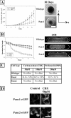
FIGURE 1.
FIGURE 1.
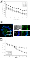
Panx1 accelerates multicellular C6 aggregate compaction. A, an in vitro 3D proliferation assay shows that 13-day growth of Panx1-expressing C6 tumor spheroids is significantly slower than that of wild-type C6 spheroids (n = 20). Scale bar = 200 μm. B, a multicellular rod compaction assay shows that Panx1-expressing C6 aggregates contract significantly faster than wild-type or Panx2 aggregates (n = 10). Scale bar = 200 μm. C, drug treatment of multicellular rods with CBX (50–100 μm) and PBN (200 μm) shows that only Panx1 rods were affected (n = 10). D, CBX treatment causes noticeable internalization of Panx1-eGFP, normally expressed uniformly on the cell membrane, whereas Panx2-eGFP is unaffected and remains intracellular. Scale bar = 20 μm.
FIGURE 2.
FIGURE 2.
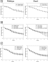
Mixing of Panx1 and Panx2 cells shows that multicellular rod contraction is proportional to the ratios of the two cell types (n = 10) (A). B, within heterocellular Panx1 (unlabeled) and Panx2 (blue) aggregates (50%:50%), localization of Panx1-eGFP and Panx2-eGFP is unchanged. Panx1 cells also compartmentalize to the center. Scale bar = 20 μm. C, C6 rods co-expressing Panx1 and Panx2 compact as rapidly as Panx1 rods (n = 10).
FIGURE 3.
FIGURE 3.
Adding exogenous ATP (500 μm) to CBX(50 μm)-treated Panx1 rods restores assembly capacity (n = 10) (A). Live (green)/dead (red) images at 24 h show that CBX+ATP-treated Panx1 aggregates resemble the control and confirm that viability was not compromised. Scale bar = 200 μm. B, wild-type and Panx1 rods show acceleration in compaction kinetics with addition of exogenous ATP alone (n = 10).
FIGURE 4.
FIGURE 4.
Panx1 rod compaction is inhibited by the nonspecific purinergic antagonist suramin (100 μm) (n = 5) (A). B, Panx1 rod compaction is accelerated by the P2X7 agonist BzATP (200–400 μm), an effect reversed with concomitant addition of the P2X7 antagonist oATP (200 μm) (n = 10). C, Panx1 rod compaction is unaffected by reagents specific for P2Y1 (agonist MeSADP, antagonist PPADS) and P2Y2 (agonist UTP) (n = 10). In all three experiments, wild-type rods were unaffected by drug treatment.
FIGURE 5.
FIGURE 5.
A. Panx1 aggregates have more robust F-actin microfilament networks (red) extending to non-cortical regions (insert). Exogenous ATP further amplifies these changes when added and can also reverse inhibitive effects of CBX treatment. Scale bar = 50 μm. B and C, only Panx1 rods are able to resume compaction upon washout of cytochalasin B (5 μm) at 3 h (▾) (n = 5), which reverses the disruptive effects of the drug on the F-actin network. Scale bar = 50 μm (B) and 20 μm (C).
FIGURE 6.
FIGURE 6.
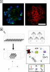
F-actin microfilament network (red) of Panx1 cells (unlabeled) is noticeably more developed than that of Panx2 cells (blue) in heterocellular aggregates (A). Scale bar = 50 μm. B, Panx1 signaling cascade and 3D multicellular aggregate compaction. Upon seeding (t0), monodispersed cells form initial cell-cell adhesive contacts via CAM interactions. This biomechanical stimulus causes Panx1 channels to open and release ATP into the extracellular space (1). ATP binds to cell surface P2X7 purinergic receptors (2), initiating a signaling cascade that increases intracellular calcium (3). These calcium waves stimulate actin microfilament organization (4), up-regulating the intercellular tensile forces that drive aggregate compaction (5).

References

    1. Sosinsky G. E., Boassa D., Dermietzel R., Duffy H. S., Laird D. W., MacVicar B. A., Naus C. C., Penuela S., Scemes E., Spray D. C., Thompson R. J., Zhao H. B., Dahl G. (2011) Pannexin channels are not gap junction hemichannels. Channels 5, 193–197 - PMC - PubMed
    1. Baranova A., Ivanov D., Petrash N., Pestova A., Skoblov M., Kelmanson I., Shagin D., Nazarenko S., Geraymovych E., Litvin O., Tiunova A., Born T. L., Usman N., Staroverov D., Lukyanov S., Panchin Y. (2004) The mammalian pannexin family is homologous to the invertebrate innexin gap junction proteins. Genomics 83, 706–716 - PubMed
    1. Penuela S., Bhalla R., Gong X. Q., Cowan K. N., Celetti S. J., Cowan B. J., Bai D., Shao Q., Laird D. W. (2007) Pannexin 1 and pannexin 3 are glycoproteins that exhibit many distinct characteristics from the connexin family of gap junction proteins. J. Cell Sci. 120, 3772–3783 - PubMed
    1. Perkins G. A., Goodenough D. A., Sosinsky G. E. (1998) Formation of the gap junction intercellular channel requires a 30 degree rotation for interdigitating two apposing connexons. J. Mol. Biol. 277, 171–177 - PubMed
    1. Bhalla-Gehi R., Penuela S., Churko J. M., Shao Q., Laird D. W. (2010) Pannexin1 and pannexin3 delivery, cell surface dynamics, and cytoskeletal interactions. J. Biol. Chem. 285, 9147–9160 - PMC - PubMed

Publication types

MeSH terms