

Esophageal squamous cell carcinoma - precursor lesions and early diagnosis
- PMID: 22267978
- PMCID: PMC3262175
- DOI: 10.4253/wjge.v4.i1.9
Esophageal squamous cell carcinoma - precursor lesions and early diagnosis
Abstract
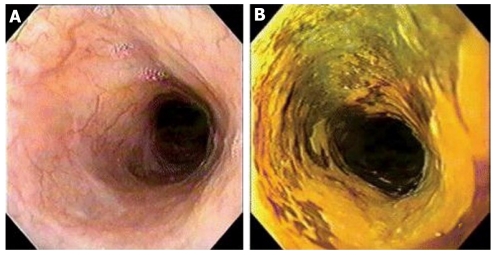
Squamous cell carcinoma of the esophagus (SCCE) carries a poor prognosis due to late diagnosis. Early detection is highly desirable, since surgical and endoscopic resection offers the only possible cure for esophageal cancer. Population screening should be undertaken in high risk areas, and in low or moderate risk areas for people with risk factors (alcoholics, smokers, mate drinkers, history of head and neck cancer, achalasia and lye stricture of the esophagus). Esophageal balloon cytology is an easy and inexpensive sampling technique, but the current methods are insufficient for primary screening due to sampling errors. Conventional endoscopy with biopsy remains the standard procedure for the identification of pre-malignant and early malignant changes in esophageal mucosa and endoscopic detection. It may be enhanced by several techniques such as dye and optic chromoendoscopy, magnifying endoscopy, and optical-based spectroscopic and imaging modalities. Since more than 80% of SCCE deaths occur in developing countries, where expensive techniques such as narrow band imaging (NBI) and autofluorescence imaging are unavailable, the most cost-effective tool for targeting biopsies may be Lugol dye chromoendoscopy, since it is easy, accurate, inexpensive and available worldwide. In ideal conditions, or in developed countries, is it reasonable to think that optimal detection will require a combination of techniques, such as the combination of Lugol's chromoendoscopy and NBI to identify esophageal areas that require further characterization by a high resolution technique. The efficacy and cost-effectiveness will determine whether these modalities will become part of standard endoscopy practice.
Keywords: Autofluorescence endoscopy; Early diagnosis; Esophageal cancer; Esophageal squamous cell carcinoma; Lugol’s solution; Narrow-band imaging endoscopy.
Figures
References
-
- Ferlay J, Shin HR, Bray F, Forman D, Mathers C, Parkin DM. Estimates of worldwide burden of cancer in 2008: GLOBOCAN 2008. Int J Cancer. 2010;127:2893–2917. - PubMed
-
- Jemal A, Bray F, Center MM, Ferlay J, Ward E, Forman D. Global cancer statistics. CA Cancer J Clin. 2010;61:69–90. - PubMed
-
- Orengo MA, Casella C, Fontana V, Filiberti R, Conio M, Rosso S, Tumino R, Crosignani P, De Lisi V, Falcini F, et al. Trends in incidence rates of oesophagus and gastric cancer in Italy by subsite and histology, 1986-1997. Eur J Gastroenterol Hepatol. 2006;18:739–746. - PubMed
LinkOut - more resources
Full Text Sources
Medical
