

doi: 10.4103/2156-7514.90482.
Epub 2011 Dec 2.
Dermatofibrosarcoma protuberans of the scalp with fibrosarcomatous degeneration and pulmonary metastasis
Affiliations
- PMID: 22267990
- PMCID: PMC3261608
- DOI: 10.4103/2156-7514.90482
Item in Clipboard
Dermatofibrosarcoma protuberans of the scalp with fibrosarcomatous degeneration and pulmonary metastasis
J Clin Imaging Sci.
2011.
Abstract
Dermatofibrosarcoma protuberans is a rare locally aggressive cutaneous tumor of intermediate malignancy. It is a slow-growing neoplasm with a marked propensity to recur after resection. Head and neck involvement is unusual and distant metastases are quite rare but tend to be more frequent in tumors that undergo fibrosarcomatous degeneration. We present the imaging and corresponding histopathology in a case of dermatofibrosarcoma protuberans of the scalp demonstrating fibrosarcomatous degeneration and lung metastasis.
Keywords: Dermatofibrosarcoma protuberans; imaging characteristics; lung metastasis; scalp.
Conflict of interest statement
Figures

27-year -ld with DFSP. (a) Axial T1 pre-contrast image demonstrates a large exophytic mass slightly hypointense to gray matter which has recurred within the left frontal scalp. (b) The mass is predominantly T2 hyperintense and demonstrates prominent heterogeneous enhancement after gadolinium on (c) axial and (d) coronal T1 post-contrast images. Areas of central necrosis or hemorrhage are noted.

27-year-old with DFSP. (a) Patient photograph and (b) corresponding saggital T1 post-contrast MRI demonstrating the recurrent DFSP which is larger, and more ulcerated than previously with continued heterogeneous enhancement.

27-year-old with DFSP. (a) Axial and (b) coronal post-contrast CT images of the chest demonstrate a large round mass with mild enhancement in the right lung centered in the right middle lobe and causing mass effect on the adjacent right atrium.
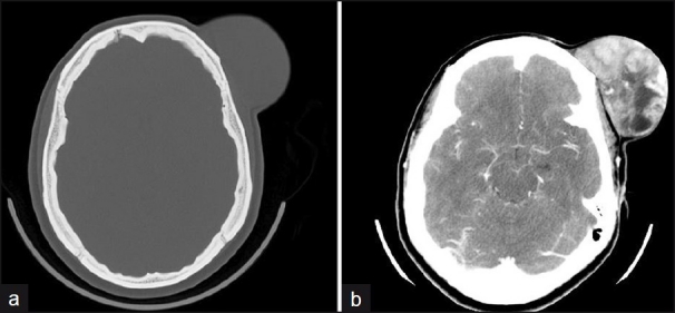
27-year-old with DFSP. Axial CT head (a) without and (b) with contrast demonstrate a large protuberant soft tissue mass arising from the left frontal scalp. The mass enhances heterogeneously after contrast with areas of central hypovascularity but no invasion of the calvarium.

27-year-old with DFSP. (a) H and E stain demonstrating spindle-type cells with areas of classic “storiform” or “cart wheel” pattern. (b) Immunostain for CD34 shows predominantly strong positivity but areas with loss of CD34 immunopositivity representing fibrosarcomatous transformation are also present (arrow).
References
-
- Darier S, Ferrand M. Dermatofibrosarcomes progressives et ricidivantes on fibrosarcomes de la peau. Ann Dermatol Venereol. 1924;5:545–62.
-
- Kransdorf MJ, Meis-Kindblom JM. Dermatofibrosarcoma protuberans: Radiologic appearance. AJR Am J Roentgenol. 1994;162:391–4. - PubMed
-
- Torreggiani WC, Al-Ismail K, Munk PL, Nicolaou S, O'Connell JX, Knowling MA. Dermatofibrosarcoma Protuberans: MR Imaging Features. AJR Am J Roentgenol. 2002;178:989–93. - PubMed
-
- Bowne WB, Antonescu CR, Leung DH, Katz SC, Hawkins WG, Woodruff JM, et al. Dermatofibrosarcoma protuberans: A clinicopathologic analysis of patients treated and followed at a single institution. Cancer. 2000;88:2711–20. - PubMed
-
- Uematsu Y, Fukai J, Tamura M, Owai Y, Obayashi S, Nakai K, et al. Distant metastasis of dermatofibrosarcoma of the scalp. Neurol Med Chir (Tokyo) 2003;43:493–6. - PubMed
LinkOut - more resources
Full Text Sources
