Imaging characteristics of oligodendrogliomas that predict grade
- PMID: 22268087
- PMCID: PMC3404805
- DOI: 10.3174/ajnr.A2895
Imaging characteristics of oligodendrogliomas that predict grade
Abstract
Background and purpose: Oligodendrogliomas are tumors that have variable WHO grades depending on anaplasia and astrocytic components and their treatment may differ accordingly. Our aim was to retrospectively evaluate imaging features of oligodendrogliomas that predict tumor grade.
Materials and methods: The imaging studies of 75 patients with oligodendrogliomas were retrospectively reviewed and compared with the histologic grade. The presence and degree of enhancement and calcification were evaluated subjectively. rCBV and ADC maps were measured. Logistic linear regression models were used to determine the relationship between imaging factors and tumor grade.
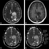
Results: Thirty of 75 (40%) tumors enhanced, including 9 of 46 (19.6%) grade II and 21 of 29 (72.4%) grade III tumors (P < .001). Grade III tumors showed lower ADC values compared with grade II tumors (odds ratio of a tumor being grade III rather than grade II = 0.07; 95% CI, 0.02-0.25; P = .001). An optimal ADC cutoff of 925 10(-6) mm(2)/s was established, which yielded a specificity of 89.1%, sensitivity of 62.1%, and accuracy of 78.7%. There was no statistically significant association between tumor grade and the presence of calcification and perfusion values. Multivariable prediction rules were applied for ADC < 925 10(-6) mm(2)/s, the presence of enhancement, and the presence of calcification. If either ADC < 925 10(-6) mm(2)/s or enhancement was present, it yielded 93.1% sensitivity, 73.9% specificity, and 81.3% accuracy. The most accurate (82.2%) predictive rule was seen when either ADC < 925 10(-6) mm(2)/s or enhancement and calcification were present.
Conclusions: Models based on contrast enhancement, calcification, and ADC values can assist in predicting the grade of oligodendrogliomas and help direct biopsy sites, raise suspicion of sampling error, and predict prognosis.
Figures


Comment in
-
[Oligodendrogliomas - MRI can assist grading].Rofo. 2013 Jun;185(6):528-9. doi: 10.1055/s-0032-1319453. Epub 2013 Jun 5. Rofo. 2013. PMID: 23740709 German. No abstract available.
References
-
- Claus EB, Black PM. Survival rates and patterns of care for patients diagnosed with supratentorial low-grade gliomas: data from the SEER program, 1973–2001. Cancer 2006;106:1358–63 - PubMed
-
- McCarthy BJ, Propp JM, Davis FG, et al. Time trends in oligodendroglial and astrocytic tumor incidence. Neuroepidemiology 2008;30:34–44 - PubMed
-
- Koeller KK, Rushing EJ. From the archives of the AFIP: Oligodendroglioma and its variants: radiologic-pathologic correlation. Radiographics 2005;25:1669–88 - PubMed
-
- Shaw EG, Scheithauer BW, O'Fallon JR, et al. Oligodendrogliomas: the Mayo Clinic experience. J Neurosurg 1992;76:428–34 - PubMed
-
- Engelhard HH, Stelea A, Mundt A. Oligodendroglioma and anaplastic oligodendroglioma: clinical features, treatment, and prognosis. Surg Neurol 2003;60:443–56 - PubMed
MeSH terms
Grants and funding
LinkOut - more resources
Full Text Sources
Medical
