

Angiographic CT with intravenous contrast injection compared with conventional rotational angiography in the diagnostic work-up of cerebral aneurysms
- PMID: 22268091
- PMCID: PMC7968818
- DOI: 10.3174/ajnr.A2883
Angiographic CT with intravenous contrast injection compared with conventional rotational angiography in the diagnostic work-up of cerebral aneurysms
Abstract
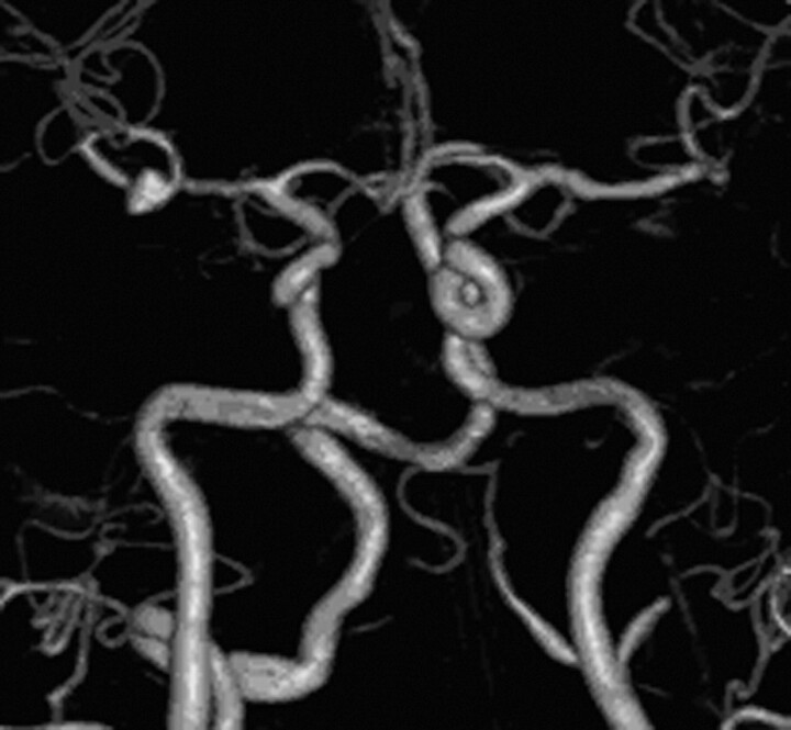
Background and purpose: Noninvasive imaging of cerebral aneurysms is still considered inferior to conventional angiography. The purpose of this study was to evaluate the diagnostic accuracy of ivACT in the assessment of intracranial aneurysms compared with 3D-DSA.
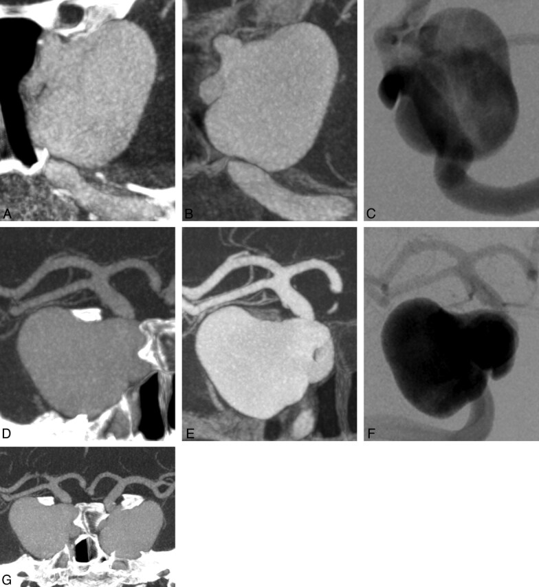
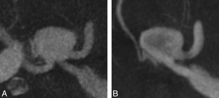
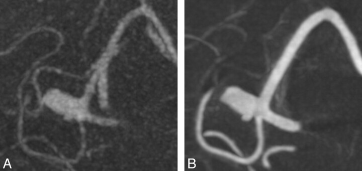
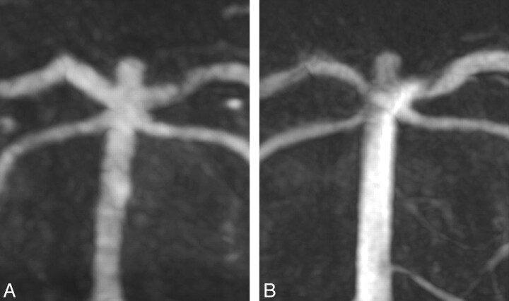
Materials and methods: We included 13 patients with 15 incidental unruptured saccular aneurysms scheduled for diagnostic angiographic work-up in our study. In each patient, we performed an ivACT and a conventional angiography including a 3D rotational run. During postprocessing, MPR images were generated for each technique. Maximal aneurysm diameter, neck diameter, aneurysm height, maximum width, bulge height, parent artery diameter, and angle between the parent artery and aneurysm apex were measured for each aneurysm.
Results: 3D-DSA and ivACT both provided images of high quality without artificial disturbances (ie, motion artifacts). Measurements of all parameters resulted in comparable values for both modalities with a strong correlation (P ≤ .001).
Conclusions: ivACT is feasible for the noninvasive visualization of saccular cerebral aneurysms and may provide reliable diagnostic information for the assessment of aneurysm size and geometry comparable with conventional intra-arterial 3D rotational angiography. These preliminary results might be a first promising step to replacing conventional angiography in preinterventional aneurysm imaging.
Figures
References
-
- Campi A, Ramzi N, Molyneux AJ, et al. Retreatment of ruptured cerebral aneurysms in patients randomized by coiling or clipping in the International Subarachnoid Aneurysm Trial (ISAT). Stroke 2007;38:1538–44 - PubMed
-
- Molyneux AJ, Kerr RS, Birks J, et al. Risk of recurrent subarachnoid haemorrhage, death, or dependence and standardised mortality ratios after clipping or coiling of an intracranial aneurysm in the International Subarachnoid Aneurysm Trial (ISAT): long-term follow-up. Lancet Neurol 2009;8:427–33 - PMC - PubMed
-
- Schievink WI, Torres VE, Piepgras DG, et al. Saccular intracranial aneurysms in autosomal dominant polycystic kidney disease. J Am Soc Nephrol 1992;3:88–95 - PubMed
-
- Brisman JL, Song JK, Newell DW. Cerebral aneurysms. N Engl J Med 2006;355:928–39 - PubMed
-
- Anxionnat R, Bracard S, Ducrocq X, et al. Intracranial aneurysms: clinical value of 3D digital subtraction angiography in the therapeutic decision and endovascular treatment. Radiology 2001;218:799–808 - PubMed
Publication types
MeSH terms
Substances
LinkOut - more resources
Full Text Sources
Medical
Miscellaneous