

Detection of intratumoral calcification in oligodendrogliomas by susceptibility-weighted MR imaging
- PMID: 22268093
- PMCID: PMC7968798
- DOI: 10.3174/ajnr.A2862
Detection of intratumoral calcification in oligodendrogliomas by susceptibility-weighted MR imaging
Abstract
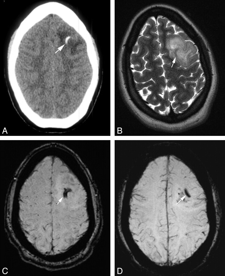
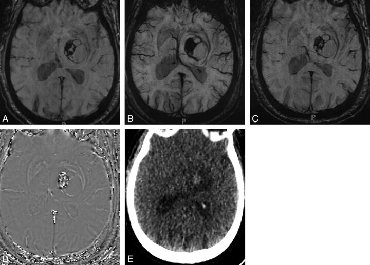
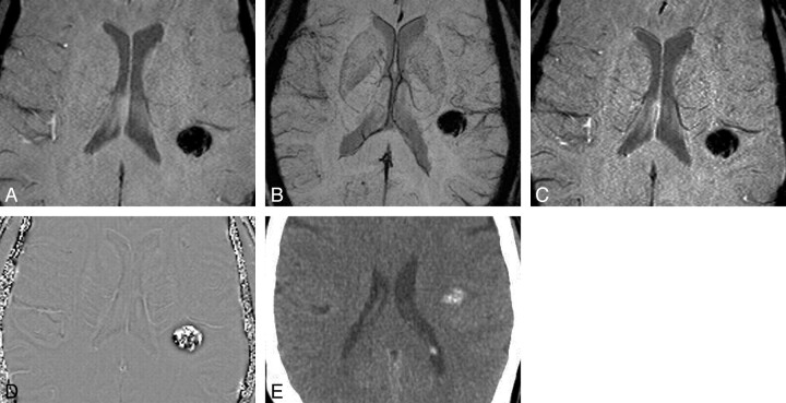
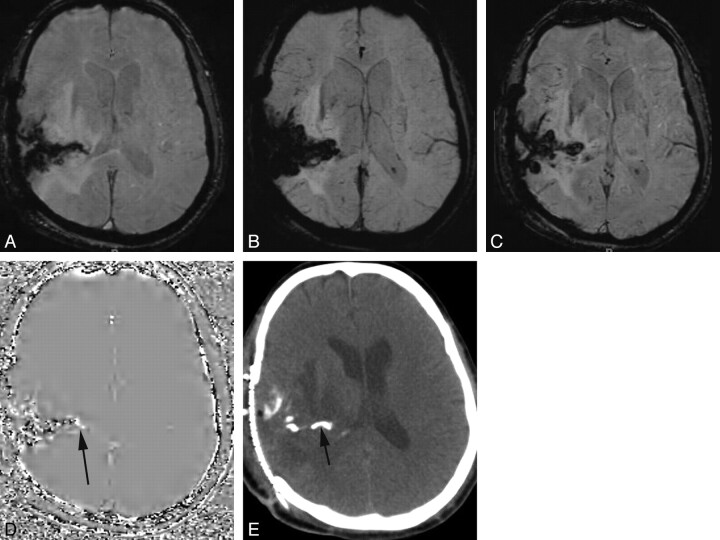
Background and purpose: SWI is a unique pulse sequence sensitive to both hemorrhage and calcification. Our aim was to retrospectively assess the ability of SWI to detect intratumoral calcification in ODs compared with conventional MR imaging.
Materials and methods: Using CT as criterion standard, the MR imaging findings from 71 patients (33 males, 38 females; mean age, 42.5 years) with pathologically proved OD were retrospectively evaluated. We classified the MR imaging data into SWI data (MRSWI) and traditional pulse sequences (MRnoSWI). The sensitivity and specificity of the MRnoSWI (n = 71) were compared with that of the MRSWI (n = 13) independently and also for matched-paired data (n = 13). The Fisher exact test was applied to the matched-pair data for statistical evaluation.
Results: For paired data of MRSWI and MRnoSWI (n = 13), there was significantly increased sensitivity of MRSWI (86%) for the detection of intratumoral calcification in OD compared with the MRnoSWI (14.3%) (P = .015, Fisher exact test) by using CT as the criterion standard. The overall accuracy of MRSWI for the paired data was also significantly greater (P = .048). The specificities were not significantly different (P = .773). The sensitivity of MRSWI (n = 13) was 86%, and for MRnoSWI (n = 71), it was 33.3%. Specificity of MRSWI was 83%, and for MRnoSWI, it was 95%.
Conclusions: SWI is better able to detect calcification in ODs than conventional MR imaging pulse sequences.
Figures
Comment in
-
High-pass-filtered phase image: left- versus right-handed MR imaging systems.AJNR Am J Neuroradiol. 2013 Jun-Jul;34(6):E72. doi: 10.3174/ajnr.A3571. Epub 2013 Mar 28. AJNR Am J Neuroradiol. 2013. PMID: 23538416 Free PMC article. No abstract available.
References
-
- Haacke EM, Cheng NY, House MJ, et al. . Imaging iron stores in the brain using magnetic resonance imaging. Magn Reson Imaging 2005;23:1–25 - PubMed
-
- Gupta RK, Rao SB, Jain R, et al. . Differentiation of calcification from chronic hemorrhage with corrected gradient echo phase imaging. J Comput Assist Tomogr 2001;25:698–704 - PubMed
-
- Margain D, Peretti-Viton P, Perez-Castillo AM, et al. . Oligodendrogliomas. J Neuroradiol 1991;18:153–60 - PubMed
MeSH terms
LinkOut - more resources
Full Text Sources
Medical