

Intramural optical mapping of V(m) and Ca(i)2+ during long-duration ventricular fibrillation in canine hearts
- PMID: 22268104
- PMCID: PMC3311483
- DOI: 10.1152/ajpheart.00426.2011
Intramural optical mapping of V(m) and Ca(i)2+ during long-duration ventricular fibrillation in canine hearts
Abstract
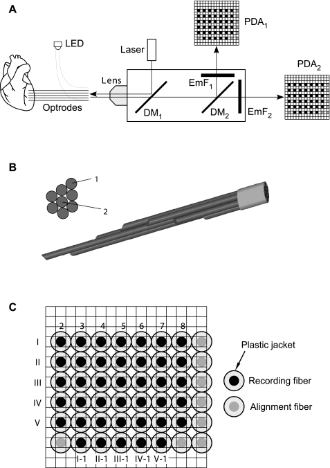
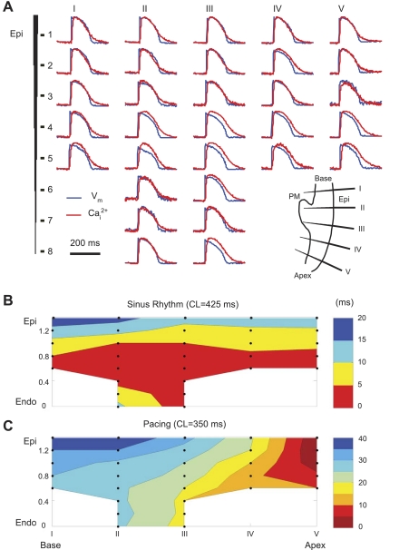
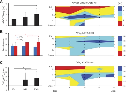
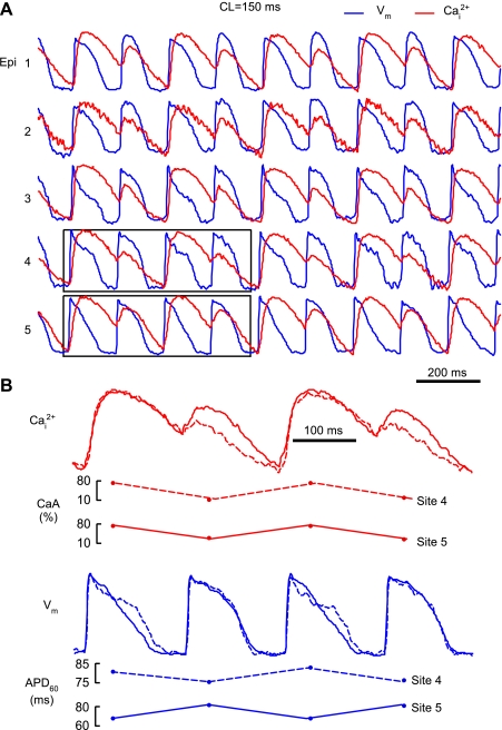
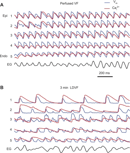
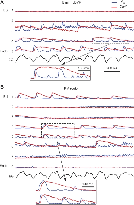
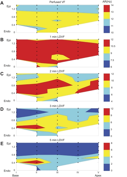
Intramural gradients of intracellular Ca(2+) (Ca(i)(2+)) Ca(i)(2+) handling, Ca(i)(2+) oscillations, and Ca(i)(2+) transient (CaT) alternans may be important in long-duration ventricular fibrillation (LDVF). However, previous studies of Ca(i)(2+) handling have been limited to recordings from the heart surface during short-duration ventricular fibrillation. To examine whether abnormalities of intramural Ca(i)(2+) handling contribute to LDVF, we measured membrane voltage (V(m)) and Ca(i)(2+) during pacing and LDVF in six perfused canine hearts using five eight-fiber optrodes. Measurements were grouped into epicardial, midwall, and endocardial layers. We found that during pacing at 350-ms cycle length, CaT duration was slightly longer (by ≃10%) in endocardial layers than in epicardial layers, whereas action potential duration (APD) exhibited no difference. Rapid pacing at 150-ms cycle length caused alternans in both APD (APD-ALT) and CaT amplitude (CaA-ALT) without significant transmural differences. For 93% of optrode recordings, CaA-ALT was transmurally concordant, whereas APD-ALT was either concordant (36%) or discordant (54%), suggesting that APD-ALT was not caused by CaA-ALT. During LDVF, V(m) and Ca(i)(2+) progressively desynchronized when not every action potential was followed by a CaT. Such desynchronization developed faster in the epicardium than in the other layers. In addition, CaT duration strongly increased (by ∼240% at 5 min of LDVF), whereas APD shortened (by ∼17%). CaT rises always followed V(m) upstrokes during pacing and LDVF. In conclusion, the fact that V(m) upstrokes always preceded CaTs indicates that spontaneous Ca(i)(2+) oscillations in the working myocardium were not likely the reason for LDVF maintenance. Strong V(m)-Ca(i)(2+) desynchronization and the occurrence of long CaTs during LDVF indicate severely impaired Ca(i)(2+) handling and may potentially contribute to LDVF maintenance.
Figures
Similar articles
-
Transmural optical measurements of Vm dynamics during long-duration ventricular fibrillation in canine hearts.Heart Rhythm. 2009 Jun;6(6):796-802. doi: 10.1016/j.hrthm.2009.02.028. Epub 2009 Feb 24. Heart Rhythm. 2009. PMID: 19467507 Free PMC article.
-
The importance of Purkinje activation in long duration ventricular fibrillation.J Am Heart Assoc. 2014 Feb 28;3(1):e000495. doi: 10.1161/JAHA.113.000495. J Am Heart Assoc. 2014. PMID: 24584738 Free PMC article.
-
Optical mapping of sarcoplasmic reticulum Ca2+ in the intact heart: ryanodine receptor refractoriness during alternans and fibrillation.Circ Res. 2014 Apr 25;114(9):1410-21. doi: 10.1161/CIRCRESAHA.114.302505. Epub 2014 Feb 25. Circ Res. 2014. PMID: 24568740 Free PMC article.
-
Developing a novel comprehensive framework for the investigation of cellular and whole heart electrophysiology in the in situ human heart: historical perspectives, current progress and future prospects.Prog Biophys Mol Biol. 2014 Aug;115(2-3):252-60. doi: 10.1016/j.pbiomolbio.2014.06.004. Epub 2014 Jun 24. Prog Biophys Mol Biol. 2014. PMID: 24972083 Review.
-
Optical and electrical recordings from isolated coronary-perfused ventricular wedge preparations.J Mol Cell Cardiol. 2013 Jan;54:53-64. doi: 10.1016/j.yjmcc.2012.10.017. Epub 2012 Nov 9. J Mol Cell Cardiol. 2013. PMID: 23142540 Free PMC article. Review.
Cited by
-
Microenvironmental Modulation of Calcium Wave Propagation Velocity in Engineered Cardiac Tissues.Cell Mol Bioeng. 2018 Apr 17;11(5):337-352. doi: 10.1007/s12195-018-0522-2. eCollection 2018 Oct. Cell Mol Bioeng. 2018. PMID: 31719889 Free PMC article.
-
Mapping cardiac surface mechanics with structured light imaging.Am J Physiol Heart Circ Physiol. 2012 Sep 15;303(6):H712-20. doi: 10.1152/ajpheart.00269.2012. Epub 2012 Jul 13. Am J Physiol Heart Circ Physiol. 2012. PMID: 22796539 Free PMC article.
-
Toward microendoscopy-inspired cardiac optogenetics in vivo: technical overview and perspective.J Biomed Opt. 2014 Aug;19(8):080701. doi: 10.1117/1.JBO.19.8.080701. J Biomed Opt. 2014. PMID: 25117076 Free PMC article. Review.
-
Cardiac optogenetics.Am J Physiol Heart Circ Physiol. 2013 May;304(9):H1179-91. doi: 10.1152/ajpheart.00432.2012. Epub 2013 Mar 1. Am J Physiol Heart Circ Physiol. 2013. PMID: 23457014 Free PMC article. Review.
-
A century of optocardiography.IEEE Rev Biomed Eng. 2014;7:115-25. doi: 10.1109/RBME.2013.2286296. Epub 2013 Oct 23. IEEE Rev Biomed Eng. 2014. PMID: 24158521 Free PMC article. Review.
References
-
- Akar FG, Yan GX, Antzelevitch C, Rosenbaum DS. Unique topographical distribution of M cells underlies reentrant mechanism of torsade de pointes in the long-QT syndrome. Circulation 105: 1247–1253, 2002 - PubMed
-
- Burashnikov A, Antzelevitch C. Reinduction of atrial fibrillation immediately after termination of the arrhythmia is mediated by late phase 3 early afterdepolarization-induced triggered activity. Circulation 107: 2355–2360, 2003 - PubMed
-
- Capogrossi MC, Houser SR, Bahinski A, Lakatta EG. Synchronous occurrence of spontaneous localized calcium release from the sarcoplasmic reticulum generates action potentials in rat cardiac ventricular myocytes at normal resting membrane potential. Circ Res 61: 498–503, 1987 - PubMed
-
- Clusin WT. Mechanisms of calcium transient and action potential alternans in cardiac cells and tissues. Am J Physiol Heart Circ Physiol 294: H1–H10, 2008 - PubMed
Publication types
MeSH terms
Substances
Grants and funding
LinkOut - more resources
Full Text Sources
Other Literature Sources
Miscellaneous