. 2012 Apr 1;185(7):744-55.
doi: 10.1164/rccm.201109-1686OC. Epub 2012 Jan 20.

Calpastatin controls polymicrobial sepsis by limiting procoagulant microparticle release

Affiliations

Calpastatin controls polymicrobial sepsis by limiting procoagulant microparticle release

Lara Zafrani et al. Am J Respir Crit Care Med. .

Abstract

Rationale: Sepsis, a leading cause of death worldwide, involves widespread activation of inflammation, massive activation of coagulation, and lymphocyte apoptosis. Calpains, calcium-activated cysteine proteases, have been shown to increase inflammatory reactions and lymphocyte apoptosis. Moreover, calpain plays an essential role in microparticle release.

Objectives: We investigated the contribution of calpain in eliciting tissue damage during sepsis.

Methods: To test our hypothesis, we induced polymicrobial sepsis by cecal ligation and puncture in wild-type (WT) mice and transgenic mice expressing high levels of calpastatin, a calpain-specific inhibitor.

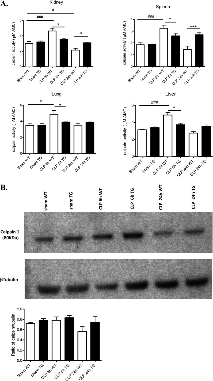
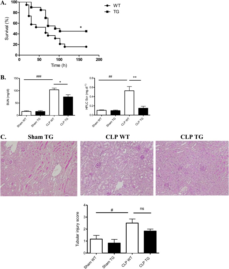
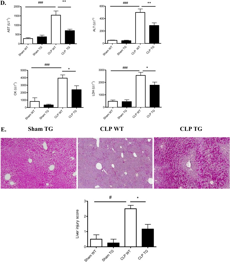
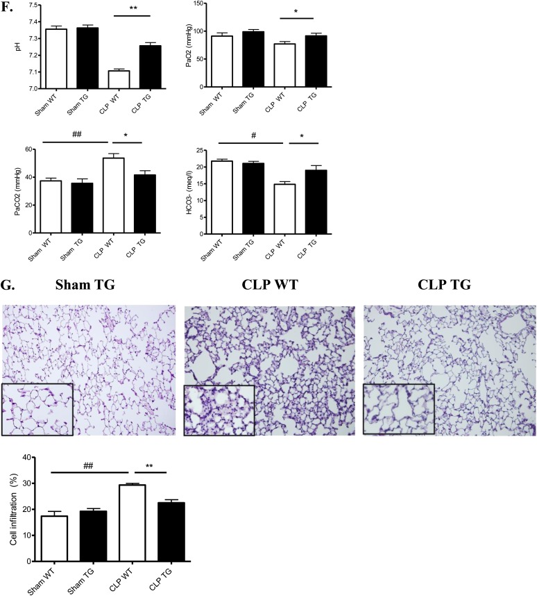
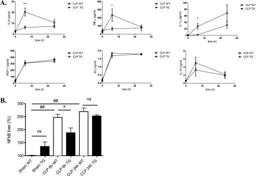
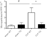
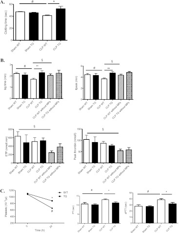
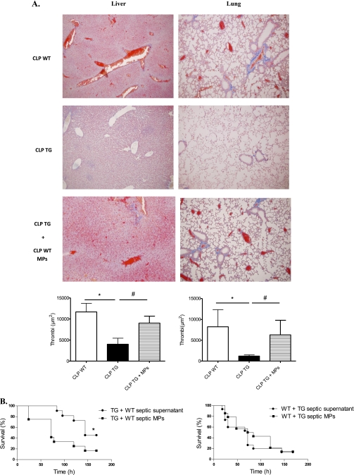
Measurements and main results: In WT mice, calpain activity increased transiently peaking at 6 hours after cecal ligation and puncture surgery. Calpastatin overexpression improved survival, organ dysfunction (including lung, kidney, and liver damage), and lymphocyte apoptosis. It decreased the sepsis-induced systemic proinflammatory response and disseminated intravascular coagulation, by reducing the number of procoagulant circulating microparticles and therefore delaying thrombin generation. The deleterious effect of microparticles in this model was confirmed by transferring microparticles from septic WT to septic transgenic mice, worsening their survival and coagulopathy.

Conclusions: These results demonstrate an important role of the calpain/calpastatin system in coagulation/inflammation pathways during sepsis, because calpain inhibition is associated with less severe disseminated intravascular coagulation and better overall outcomes in sepsis.


Figures

Figure 1.
Figure 1.
Alterations of calpain activity and expression in tissues of wild-type (WT) and transgenic (TG) mice following cecal ligation and puncture (CLP). (A) Kidney, liver, spleen, and lung were isolated from WT (white bars) and TG (black bars) mice 6 and 24 hours after CLP (n = 5 mice per group). Calpain activities were determined by measuring the calpain-specific cleavage of fluorescent N-succinyl-Leu-Tyr-amidomethylcoumarin, a calpain-specific substrate. (B) Western blot analysis of calpain 1 in kidney from WT and TG before and 6 and 24 hours after CLP. Quantification was done by densitometry (n = 3). *P < 0.05 WT versus TG, #P < 0.05, ##P < 0.01, ###P < 0.001 CLP versus sham. Error bars represent means ± SEM.
Figure 2.
Figure 2.
Effect of calpastatin overexpression on survival and multiple organ failure during sepsis. (A) Survival curves of wild-type (WT) and transgenic (TG) mice after cecal ligation and puncture (CLP) (P ≤ 0.02). (B) Calpastatin overexpression effect on kidney function, as reflected by serum creatinine concentration (HPLC SCr) and serum urea (BUN) in sham and septic mice 24 hours after CLP (n = 8 in each sham group and 14 in each septic group). (C) Representative periodic acid Schiff staining in kidney of sham TG, septic WT, and TG mice (original magnification, ×20) and tubular injury scores 24 hours after CLP (n = 4 in sham group and 6 in septic group). (D) Alanine aminotransferase (ALT), aspartate aminotransferase (AST), creatine kinase (CK), and lactate dehydrogenase (LDH) in sham WT, sham TG, septic WT, and septic TG mice 24 hours after CLP (n = 8 mice in each sham group and 14 mice in each septic group). (E) Representative liver histology in sham TG, septic WT, and septic TG mice (original magnification, ×10) and liver injury score 24 hours after CLP. The red staining by periodic acid Schiff in hepatocytes indicates glycogen storage capacity (n = 6 per group). (F) Arterial blood gas analysis was performed in sham and septic WT and TG mice at 24 hours (n = 6 per group). (G) Hematoxylin and eosin staining was performed on lung sections from sham TG, septic WT, and septic TG mice at 24 hours (×20 and ×40) and morphometric analysis of cell infiltration was performed using an automated analysis system (n = 12 per group). *P < 0.05 WT versus TG, **P < 0.01 WT versus TG, #P < 0.05 CLP versus sham, ##P < 0.01 CLP versus sham, ###P < 0.001 CLP versus sham. Error bars represent means ± SEM. ns = nonsignificant.
Figure 2.
Figure 2.
Effect of calpastatin overexpression on survival and multiple organ failure during sepsis. (A) Survival curves of wild-type (WT) and transgenic (TG) mice after cecal ligation and puncture (CLP) (P ≤ 0.02). (B) Calpastatin overexpression effect on kidney function, as reflected by serum creatinine concentration (HPLC SCr) and serum urea (BUN) in sham and septic mice 24 hours after CLP (n = 8 in each sham group and 14 in each septic group). (C) Representative periodic acid Schiff staining in kidney of sham TG, septic WT, and TG mice (original magnification, ×20) and tubular injury scores 24 hours after CLP (n = 4 in sham group and 6 in septic group). (D) Alanine aminotransferase (ALT), aspartate aminotransferase (AST), creatine kinase (CK), and lactate dehydrogenase (LDH) in sham WT, sham TG, septic WT, and septic TG mice 24 hours after CLP (n = 8 mice in each sham group and 14 mice in each septic group). (E) Representative liver histology in sham TG, septic WT, and septic TG mice (original magnification, ×10) and liver injury score 24 hours after CLP. The red staining by periodic acid Schiff in hepatocytes indicates glycogen storage capacity (n = 6 per group). (F) Arterial blood gas analysis was performed in sham and septic WT and TG mice at 24 hours (n = 6 per group). (G) Hematoxylin and eosin staining was performed on lung sections from sham TG, septic WT, and septic TG mice at 24 hours (×20 and ×40) and morphometric analysis of cell infiltration was performed using an automated analysis system (n = 12 per group). *P < 0.05 WT versus TG, **P < 0.01 WT versus TG, #P < 0.05 CLP versus sham, ##P < 0.01 CLP versus sham, ###P < 0.001 CLP versus sham. Error bars represent means ± SEM. ns = nonsignificant.
Figure 3.
Figure 3.
Effect of calpastatin overexpression on cytokine concentrations and nuclear factor (NF)-κB activity. (A) Serum interleukin (IL)-6, tumor necrosis factor α(TNFα), IL-1α, keratinocyte-derived chemokine (KC), monocyte chemotactic protein (MCP)-1, and IL-10 concentrations 6 and 24 hours after cecal ligation and puncture (CLP) in wild-type (WT) and transgenic (TG) mice measured by ELISA (n = 4–6 mice per group). (B) NF-κB nuclear activity in liver of sham and septic mice, WT and TG, at 6 and 24 hours (n = 4 mice per group). Error bars represent means ± SEM; *P < 0.05 WT versus TG, ***P < 0.001 WT versus TG, ##P < 0.01 CLP versus sham. ns = nonsignificant.
Figure 4.
Figure 4.
Flow cytometry data on circulating microparticles (MPs) in sham and septic mice at 24 hours after cecal ligation and puncture (CLP) surgery. Data are given as absolute count of MPs per milliliter of platelet-free plasma (PFP) · 106 (n = 6 per group). Error bars represent means ± SEM; *P < 0.05 WT versus CLP TG, #P < 0.05 CLP versus sham.
Figure 5.
Figure 5.
Procoagulant activity of microparticles (MPs). Effect of calpastatin overexpression on thrombin generation and global test of hemostasis. (A) Procoagulant activity of MPs was assessed by functional assay (STA-Procoag-PPL, Diagnostica Stago) in sham wild-type (WT), sham transgenic (TG), septic WT, and septic TG mice 24 hours after cecal ligation and puncture (CLP) surgery (n = 8 per group) (P ≤ 0.04). Error bars represent means ± SEM; *P < 0.05 WT versus TG, #P < 0.05 CLP versus sham. (B) Calibrated thrombography in PPP samples from sham WT, sham TG, septic WT, and septic TG mice and PPP depleted in MPs from CLP WT and CLP TG mice, 24 hours after CLP surgery. The prolongation of the lag time and time to peak thrombin generation suggest a delay in the activation of hemostasis in TG mice. **P < 0.01 versus CLP WT; #P < 0.05 versus sham WT; §P < 0.05 CLP WT versus PPP depleted in MPs. Results are expressed as mean (±SEM). (C) Platelets and hemostatic parameters. Measurement of platelets are shown before and 24 hours after CLP surgery (n = 5 per group). Prothrombin time (PT) and activated partial thromboplastin time (aPTT) are shown in plasma from sham WT, sham TG, septic WT, and septic TG mice, 24 hours after CLP surgery. PT and aPTT are significantly increased in septic WT mice (n = 5 per group). *P < 0.05, versus CLP WT; #P < 0.05 versus sham WT. Results are expressed as mean (±SEM).
Figure 6.
Figure 6.
Effect of microparticles (MPs) on fibrin deposit and survival. (A) Representative photographs of sections stained with Marcius Scarlet Blue for fibrin. Results shown correspond to livers (original magnification, ×20) and lungs (original magnification, ×10) in “pre-mortem” mice. Thrombi (red blood cells mixed with fibrin in red) were seen in wild-type (WT) septic mice and transgenic (TG) mice supplemented with WT septic MPs. Quantification of red area was performed using an automated analysis system (n = 6 per group). Results are means ± SEM. *P < 0.05 versus WT, #P < 0.05 CLP TG + MPs versus CLP TG. (B) Effects of MPs on survival. (Left) Survival curves of septic TG mice supplemented with MPs from septic WT mice (n = 16) or corresponding supernatant (n = 14) (P ≤ 0.04); (right) survival curves of septic WT mice supplemented with MPs from septic TG mice (n = 12) or corresponding supernatant (n = 12) (P = nonsignificant). CLP = cecal ligation and puncture.

References

    1. Vincent JL, Rello J, Marshall J, Silva E, Anzueto A, Martin CD, Moreno R, Lipman J, Gomersall C, Sakr Y, et al. International study of the prevalence and outcomes of infection in intensive care units. JAMA 2009;302:2323–2329 - PubMed
    1. Levy MM, Fink MP, Marshall JC, Abraham E, Angus D, Cook D, Cohen J, Opal SM, Vincent JL, Ramsay G. 2001 SCCM/ESICM/ACCP/ATS/SIS International Sepsis Definitions Conference. Crit Care Med 2003;31:1250–1256 - PubMed
    1. Annane D, Bellissant E, Cavaillon JM. Septic shock. Lancet 2005;365:63–78 - PubMed
    1. Esmon CT. Sepsis: a myriad of responses. Lancet 2001;358:S61. - PubMed
    1. Morel O, Toti F, Hugel B, Freyssinet JM. Cellular microparticles: a disseminated storage pool of bioactive vascular effectors. Curr Opin Hematol 2004;11:156–164 - PubMed

Publication types