. 2012 Sep;16(9):2176-85.
doi: 10.1111/j.1582-4934.2012.01526.x.

Beneficial effects of edaravone, a novel antioxidant, in rats with dilated cardiomyopathy

Affiliations

Beneficial effects of edaravone, a novel antioxidant, in rats with dilated cardiomyopathy

Somasundaram Arumugam et al. J Cell Mol Med. 2012 Sep.

Abstract

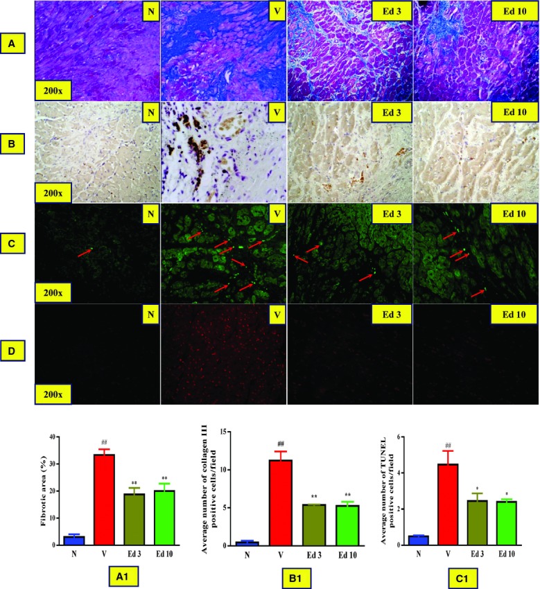
Edaravone, a novel antioxidant, acts by trapping hydroxyl radicals, quenching active oxygen and so on. Its cardioprotective activity against experimental autoimmune myocarditis (EAM) was reported. Nevertheless, it remains to be determined whether edaravone protects against cardiac remodelling in dilated cardiomyopathy (DCM). The present study was undertaken to assess whether edaravone attenuates myocardial fibrosis, and examine the effect of edaravone on cardiac function in rats with DCM after EAM. Rat model of EAM was prepared by injection with porcine cardiac myosin 28 days after immunization, we administered edaravone intraperitoneally at 3 and 10 mg/kg/day to rats for 28 days. The results were compared with vehicle-treated rats with DCM. Cardiac function, by haemodynamic and echocardiographic study and histopathology were performed. Left ventricular (LV) expression of NADPH oxidase subunits (p47(phox), p67(phox), gp91(phox) and Nox4), fibrosis markers (TGF-β(1) and OPN), endoplasmic reticulum (ER) stress markers (GRP78 and GADD 153) and apoptosis markers (cytochrome C and caspase-3) were measured by Western blotting. Edaravone-treated DCM rats showed better cardiac function compared with those of the vehicle-treated rats. In addition, LV expressions of NADPH oxidase subunits levels were significantly down-regulated in edaravone-treated rats. Furthermore, the number of collagen-III positive cells in the myocardium of edaravone-treated rats was lower compared with those of the vehicle-treated rats. Our results suggest that edaravone ameliorated the progression of DCM by modulating oxidative and ER stress-mediated myocardial apoptosis and fibrosis.


Figures

Fig 1
Fig 1
Echocardiographic data. Group N, age-matched intact rats; Group V, rats with dilated cardiomyopathy (DCM) treated with vehicle; Group Ed 3, rats with DCM treated with edaravone (3 mg/kg); Group Ed 10, rats with DCM treated with edaravone (10 mg/kg).
Fig 2
Fig 2
(A and A1) Azan-Mallory staining for fibrosis of the cross-sectional tissue slices of hearts (Fibrosis is indicated by blue area as opposed to the red myocardium and bar graph showing the percentage fibrotic area, respectively). (B and B1) Immunohistochemical stained myocardial tissue sections for collagen III and bar graph showing the average number of collagen-III positive cells/field, respectively. (C and C1) TUNEL stained myocardial tissue sections for apoptotic nuclei and bar graph showing the average number of TUNEL positive apoptotic nuclei/field, respectively. (D) Dihydroethidium-stained myocardial tissue sections for superoxide production. Group N: age-matched intact rats; Group V: rats with dilated cardiomyopathy (DCM) treated with vehicle; Group Ed 3: rats with DCM treated with edaravone (3 mg/kg); Group Ed 10: rats with DCM treated with edaravone (10 mg/kg); ##P < 0.01 versus Group N, *P < 0.05 and **P < 0.01 versus Group V (one-way ANOVA followed by Dunnett's test). Values are expressed as mean ± S.E.M., n = 3.
Fig 3
Fig 3
Myocardial expressions of osteopontin (OPN), transforming growth factor-β1 (TGF-β1) and cytochrome (cyt)-C. (A–C) Densitometric data of protein analysis. The mean density values of OPN, TGF-β1 and cyt-C (Expressed as a ratio relative to that of GAPDH) with representative Western blots showing specific bands for OPN, TGF-β1 and cyt-c. GAPDH was used as an internal control. Group N: age-matched intact rats; Group V: rats with dilated cardiomyopathy (DCM) treated with vehicle; Group Ed 3: rats with DCM treated with edaravone (3 mg/kg); Group Ed 10: rats with DCM treated with edaravone (10 mg/kg). ##P < 0.01 versus Group N, *P < 0.05 and **P < 0.01 versus Group V (one-way ANOVA followed by Dunnett's test). Values are expressed as mean ± S.E.M., n = 3.
Fig 4
Fig 4
Myocardial expressions of p47phox, p67phox, gp91phox and Nox4. (A–D) Densitometric data of protein analysis. The mean density values of p47phox, p67phox, gp91phox and Nox4 (Expressed as a ratio relative to that of GAPDH) with representative Western blots showing specific bands for p47phox, p67phox, gp91phox and Nox4. GAPDH was used as an internal control. An equal amount of protein sample obtained from whole ventricular homogenate was applied in each lane. These bands are representative of three separate experiments. Group N: age-matched intact rats; Group V: rats with dilated cardiomyopathy (DCM) treated with vehicle; Group Ed 3: rats with DCM treated with edaravone (3 mg/kg); Group Ed 10: rats with DCM treated with edaravone (10 mg/kg). ##P < 0.01 versus Group N, *P < 0.05 and **P < 0.01 versus Group V (one-way ANOVA followed by Dunnett's test). Values are expressed as mean ± S.E.M., n = 3.
Fig 5
Fig 5
Myocardial expressions of GRP78, GADD153, and caspase-3. (A–D) Densitometric data of protein analysis. The mean density values of GRP78, GADD153 and caspase-3 (expressed as a ratio relative to that of GAPDH) with representative Western blots showing specific bands for GRP78, GADD 153, Cyt C and caspase-3. GAPDH was used as an internal control. Group N: age-matched intact rats; Group V: rats with DCM treated with vehicle; Group Ed 3: rats with dilated cardiomyopathy (DCM) treated with edaravone (3 mg/kg); Group Ed 10: rats with DCM treated with edaravone (10 mg/kg). ##P < 0.01 versus Group N, *P < 0.05 and **P < 0.01 versus Group V (one-way ANOVA followed by Dunnett's test). Values are expressed as mean ± S.E.M., n = 3.

Similar articles

Cited by

References

    1. Kawai C. From myocarditis to cardiomyopathy: mechanisms of inflammation and cell death. Circulation. 2001;34:199–204. - PubMed
    1. Kodama M, Hanawa H, Saeki M, et al. Rat dilated cardiomyopathy after autoimmune giant cell myocarditis. Circ Res. 1994;75:278–84. - PubMed
    1. Okura Y, Yamamoto T, Goto S, et al. Characterization of cytokine and iNOS mRNA expression in situ during the course of experimental autoimmune myocarditis in rats. J Mol Cell Cardiol. 1997;29:491–502. - PubMed
    1. Ishiyama S, Hiroe M, Nishikawa T, et al. The Fas/Fas ligand system is involved in the pathogenesis of autoimmune myocarditis in rats. J Immunol. 1998;161:4695–701. - PubMed
    1. Ishiyama S, Hiroe M, Nishikawa T, et al. Nitric oxide contributes the progression of myocardial damage in experimental autoimmune myocarditis in rats. Circulation. 1997;95:489–96. - PubMed

Publication types

MeSH terms