. 2012 Jan 23:13:3.
doi: 10.1186/1471-2199-13-3.

The transcriptional activator ZNF143 is essential for normal development in zebrafish

Affiliations

The transcriptional activator ZNF143 is essential for normal development in zebrafish

Kari M Halbig et al. BMC Mol Biol. .

Abstract

Background: ZNF143 is a sequence-specific DNA-binding protein that stimulates transcription of both small RNA genes by RNA polymerase II or III, or protein-coding genes by RNA polymerase II, using separable activating domains. We describe phenotypic effects following knockdown of this protein in developing Danio rerio (zebrafish) embryos by injection of morpholino antisense oligonucleotides that target znf143 mRNA.

Results: The loss of function phenotype is pleiotropic and includes a broad array of abnormalities including defects in heart, blood, ear and midbrain hindbrain boundary. Defects are rescued by coinjection of synthetic mRNA encoding full-length ZNF143 protein, but not by protein lacking the amino-terminal activation domains. Accordingly, expression of several marker genes is affected following knockdown, including GATA-binding protein 1 (gata1), cardiac myosin light chain 2 (cmlc2) and paired box gene 2a (pax2a). The zebrafish pax2a gene proximal promoter contains two binding sites for ZNF143, and reporter gene transcription driven by this promoter in transfected cells is activated by this protein.

Conclusions: Normal development of zebrafish embryos requires ZNF143. Furthermore, the pax2a gene is probably one example of many protein-coding gene targets of ZNF143 during zebrafish development.


Figures
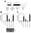
Figure 1
Figure 1
Identification of transcriptional activating domain at the amino-terminus of zebrafish ZNF143. (A) Primary structure of zebrafish ZNF143. Numbers on top depict the amino acids at the beginning and end of prominent primary structure features of the protein. Numbers in parentheses are percentages of identical amino acid residues between the zebrafish and human proteins for various regions of primary structure. (B) HEK293 cells were transfected with 200 ng of pGL3/G5AdMLT firefly luciferase reporter plasmid, 10 ng of the particular pCI/GAL4DBD/zZNF143 effector plasmid DNA noted in the figure, and 200 ng of pRL-SV40 renilla luciferase reporter. The fold-activation was determined by comparing the firefly luciferase/renilla luciferase ratios for each sample to that ratio for samples where no effector plasmid was added. Bar height shows the mean number from different transfected samples (number of experiments reported in parentheses), and the error bar represents the standard deviation from the mean. Double asterisks signify p-value <0.01, and single asterisks signify a p-value <0.05 relative to samples transfected with GAL4DBD only. The panel below shows the relative expression of each GAL4DBD/zZNF143 fusion protein in HEK293 cells by immunoblot analysis using anti-GAL4DBD antibodies. (C) Zebrafish ZF4 cells were transfected, and relative expression levels analyzed exactly as described for (B). We were unable to detect GAL4DBD/zZNF143 fusion proteins in transfected ZF4 cells, possibly because of the much lower transfection efficiency compared to HEK293 cells.
Figure 2
Figure 2
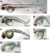
MO-induced knockdown of ZNF143 produces multiple phenotypes in zebrafish embryos. Zebrafish embryos were injected with a combination of "Start" and "5'UTR" morpholino oligonucleotides, each at 2 ng/nL. ZNF143 morphants at approximately 48 hpf are displayed according to increasing level of severity of axial development and other defects as described in the text (classified 1-5, respectively).
Figure 3
Figure 3
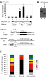
Rescue of MO phenotypes by co-injection of synthetic zebrafish ZNF143 mRNA. (A) Transcriptional activation of myc-tagged zebrafish ZNF143 deletion proteins in transfected ZF4 cells. Cells were transfected with 200 ng of firefly luciferase reporter plasmid, plus 200 ng of pRL-SV40 renilla luciferase reporter plasmid, plus various ZNF143 effector plasmids as noted in the figure at 5 ng (columns 2, 4, 6) or 25 ng (columns 3, 5, 7). Fold-activation was determined by comparing the firefly/renilla luciferase ratio for each sample to that ratio for the sample shown in column 2. Bar height shows the mean number from different experiments, and error bars report the standard deviation from the mean (when n = 3), or the range (when n = 2). Double asterisks signify a p-value <0.01, and single asterisks signify a p-value <0.05 relative to samples transfected with vector only (lane 2). (B) Relative expression of myc-tagged zebrafish ZNF143 proteins in transfected HEK293 cells. (C) Zebrafish embryos were injected with a combination of "Start" and "5'UTR" MOs along with either no RNA (MOs only), synthetic mRNA encoding wt zebrafish ZNF143 (MOs + FL), or synthetic mRNA encoding zebrafish ZNF143/Δ2-225 (MOs + Δ2-225). Embryos were scored as either dead, wt, or classes 1-6 (see Figure 2 for examples of these classifications) at 48 hpf. Results were averaged from three separate injection experiments for each condition (approximately 100 embryos per injection). The ranges in % wt phenotype for each condition in various experiments were: MOs only: 9-11%; MOs + FL: 69-72%; MOs + Δ2-225: 11-15%.
Figure 4
Figure 4
Expression analysis of ZNF143 requirement during embryonic development. In situ hybridizations to detect gata1 (A-D), cmlc2 (E-I), and pax2a (J-N) expression. (A, C, E, J, M) Wild-type. (B, D, F, G, H, I, K, L, N) ZNF143 morphants. (A, B) 16 somite stage, flat mounted embryos, dorsal view, anterior left. Brackets indicate gata1+ hematopoietic progenitors. Note reduced expression in the morphant (B). (C, D) 24 hpf, lateral views of tails. Note reduced gata1 expression in the ICM (arrows) of ZNF143 morphants (D) compared to wt (C). (E-G) approximately 27 hpf, dorsal views, anterior up. Note heart laterality in wt (E). (F, G) Two different phenotypes observed in ZNF143 morphants. While they show differences in tube morphogenesis, both lack laterality. (H, I) approximately 37 hpf, dorsal views, anterior up. Two different phenotypes in ZNF143 morphants. (J-L) 16 somite stage, flat mount, dorsal view, anterior left. Compared to the wt (J), ZNF143 morphants (K, L) display reduced pax2a expression in the pronephros (arrowheads), otic vesicles (arrows) and MHB (brackets). Note that expression in these domains is reduced in the morphants even though expression in the optic stalks is similar to wt (asterisks). (M, N) Approximately 27 hpf, dorsal views, anterior left. Expression of pax2a appears largely normal in ZNF143 morphants (N), with the exception of a substantial decrease in expression in hindbrain neurons (arrows). Scale bars: 100 μm in J-L, 200 μm in M, N.
Figure 5
Figure 5
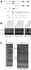
ZNF143 binds to the zebrafish pax2a promoter in vitro. (A) Map of zebrafish pax2a proximal promoter showing three potential SPH sites. The sequences of the potential SPH sites are compared to the zebrafish U6-1 element [29] and the consensus SPH sequence from [37]. Capital letters depict nucleotides matching the consensus. To facilitate binding assays two separate radiolabeled zebrafish pax2a probes were prepared by PCR as diagrammed. (B) Electrophoretic mobility shift assay. Approximately 3 fmol of radiolabeled probes depicted in (A) were incubated with 3 μL of human ZNF143-(88-638) expressed by in vitro transcription/translation (lanes 3-9; 12-18; 21-24) or with unprogrammed extract (lanes 2, 11, 20), and electrophoresed. In addition, in samples shown in lanes 4-6, 13-15, and 22-24, an unlabeled double-stranded oligonucleotide with the sequence including the human U6-1 SPH element was added with the labeled probe in amounts noted, or similarly, an unlabeled oligonucleotide containing the unrelated, OCT sequence was added to samples in lanes 7-9 and 16-18. (C) DNase I footprinting assay. Approximately 3 fmol of singly end-labeled zebrafish pax2a probe from -154 to +16 were incubated with amounts of purified human ZNF143 DBD protein as indicated. The wt pax2a promoter was used for samples in lanes 1-5, whereas SPH2 or SPH3 mutant promoters were used for samples in lanes 6-9, or lanes 10-13, respectively.
Figure 6
Figure 6
SPH sites in the zebrafish pax2a promoter are functional for transcription in transfected ZF4 cells. Zebrafish ZF4 cells were transfected per well with 200 ng plasmid DNA containing wt or SPHMUT pax2a promoters driving firefly luciferase expression, plus 200 ng pRL-SV40 renilla luciferase plasmid as a normalization control, plus 25 ng of either empty vector pCI-basic plasmid (unfilled columns) or pCI/myczZNF143 to express myc-tagged full-length ZNF143 (filled columns). The ratio of luciferase/renilla light units was determined for each sample, and these ratios were compared relative to the wt promoter with no transfected ZNF143 plasmid included in every experiment. The height of each column represents the average value for the number of independently transfected samples (specified in parentheses), and error bars depict one standard deviation above and below the mean. A double asterisk signifies a p-value <0.01, and a single asterisk signifies a p-value <0.05 comparing like reporter samples with and without addition of ZNF143 expression plasmid.

Similar articles

Cited by

References

    1. Zamrod Z, Stumph WE. U4B snRNA gene enhancer activity requires functional octamer and SPH motifs. Nucleic Acids Res. 1990;18:7323–7330. - PMC - PubMed
    1. Kunkel GR, Cheung TC, Miyake JH, Urso O, McNamara-Schroeder KJ, Stumph WE. Identification of a SPH element in the distal region of a human U6 small nuclear RNA gene promoter and characterization of the SPH binding factor in HeLa cell extracts. Gene Expr. 1996;6:59–72. - PMC - PubMed
    1. Schaub M, Myslinski E, Schuster C, Krol A, Carbon P. Staf, a promiscuous activator for enhanced transcription by RNA polymerases II and III. EMBO J. 1997;16:173–181. - PMC - PubMed
    1. Myslinski E, Krol A, Carbon P. ZNF76 and ZNF143 Are Two Human Homologs of the Transcriptional Activator Staf. J Biol Chem. 1998;273:21998–22006. - PubMed
    1. Myslinski E, Gerard MA, Krol A, Carbon P. A Genome Scale Location Analysis of Human Staf/ZNF143-binding Sites Suggests a Widespread Role for Human Staf/ZNF143 in Mammalian Promoters. J Biol Chem. 2006;281:39953–39962. - PubMed

Publication types

MeSH terms