Review
. 2012 Jan 23;5(1):2.
doi: 10.1186/1756-6606-5-2.

Understanding the physiological roles of the neuronal calcium sensor proteins

Affiliations
Review

Understanding the physiological roles of the neuronal calcium sensor proteins

Robert D Burgoyne et al. Mol Brain. .

Abstract

Calcium signalling plays a crucial role in the control of neuronal function and plasticity. Changes in neuronal Ca2+ concentration are detected by Ca2+-binding proteins that can interact with and regulate target proteins to modify their function. Members of the neuronal calcium sensor (NCS) protein family have multiple non-redundant roles in the nervous system. Here we review recent advances in the understanding of the physiological roles of the NCS proteins and the molecular basis for their specificity.


Figures
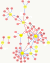
Figure 1
Figure 1
An interaction map of the known binding partners of the NCS proteins. The NCS protein family members are shown in yellow and binding partners in red with interactions directly with NCS proteins or between binding partners indicated. The edge colours indicate whether the interaction are with the Ca2+ -bound form (red) or the apo form (blue) or if this is not applicable (black). The binding partners and their abbreviations are as follows: Actin, Alsin, AP1 (clathrin adaptor), AP2 (clathrin adaptor), ARF1 (ADP-ribosylation factor-1), Calden (Caldendrin), CAPS (Ca2+-dependent activator protein for secretion), Cav1.2 (Ca2+ channel), Cav2.1 (Ca2+ channel), CALN (Calcineurin), Clathrin (Clathrin heavy chain), CREB (cAMP responsive element binding protein), CREM, (cAMP response element modulator), CTBP1/2 (C-terminal binding protein 1), cytb5 (cytochrome b5), D2R (dopamine D2 receptor), DAN (differential screening-selective gene aberrant in neuroblastome), GalT2 (galactosyl transferase 2), GluR6 (glutamate receptor 6), GRK1/2/6 (G protein-dependent receptor kinase 1/2/6), GUCY2A/2B/2D/2E (guanylate cyclase 2A/2B/2D/2E), HINT (histidine triad protein), ILIRAPL1 (interleukin receptor accessory protein like-1), IP3R (inositol 1, 4, 5 trisphosphate receptor), Kv1.5 (potassium channel), Kv4 (potassium channel), MLK2 (Mixed lineage kinase 2), NAIP (Neuronal apoptosis inhibitory protein), Nic a4b2 (nicotinic receptor containing 4 α and 2 β subunits), NR1 (NMDA receptor type 1), P2Y2R (purinergic receptor type 2Y2R), PDE (phosphodiesterase 1A), PI4KIIIb (phosphatidylinositol-4-kinase IIIβ), PICK1 (Protein interacting with C kinase 1), PINK1 (PTEN-induced kinase), prdx3 (Peroxiredoxin 3), PSD95 (post-synaptic density protein 95), PSEN1/2 (presenilin 1/2), ribeye (synaptic ribbon protein), S100b (S100 protein beta), SCAMC2 (Short calcium binding mitochondrial carrier 2), TGFbR1 (transforming growth factor β receptor type 1), TRPC1/5 (transient receptor potential channel 1/5), TSHR (thyroid stimulating hormone receptor), TTF-1 (Thyroid transcription factor 1), tubulin (α and β tubulin), ubc9 (Ubiquitin conjugating enzyme 9), VAMP2 (vesicle -associated membrane protein 2), VitDR (vitamin D receptor). Note that for simplicity we have used the name KChIP3 despite the fact that several of the original descriptions of the interactions used the alternative terminology of DREAM or calsenilin (see main text).
Figure 2
Figure 2
Structures of NCS proteins showing interactions within the exposed hydrophobic groove. (A) Structure of Ca2+-bound bovine recoverin with bound N-terminal fragment of rhodopsin kinase including residues 1-25 ([37]; PDB, 2I94). Recoverin is shown in space-filling representation and the rhodopsin kinase is shown in yellow. The rhodopsin kinase fragment forms an α-helix that is embedded in an N-terminal hydrophobic pocket of recoverin. The asterisk indicates the position of the C-terminus of recoverin. (B) Structure of Ca2+-bound human KChIP1 showing the binding of the N-terminal residues 3-27 of Kv4.3 ([143,144]; PDB, 2I2N). KChIP1 is shown in space-filling representation and the Kv4.3 fragment which forms an α-helix shown in yellow is embedded in a hydrophobic groove running across KChIP1. An additional interaction site of Kv4.3 with helix H2 of KChIP1 is omitted for clarity. The asterisk indicates the position of the C-terminus of KChIP1. (C) Structure of of Ca2+-bound mouse KChIP4a ([102];PDB, 3DD4) with the structure shown in space-filling representation apart from the N-terminal residues 1-23 which are shown in yellow. The N-terminus forms an α-helix which is embedded within the N-terminal part of the exposed hydrophobic groove of KChIP4a. The asterisk indicates the position of the C-terminus of KChIP4a. (D) Structure of of Ca2+-bound S. cerevisiae Frq1 with bound Pik1(121-175) ([142];PDB, 2JU0) the structure of Frq1 is shown in space-filling representation and the Pik1 fragment in yellow. Pik1 forms two α-helices joined by a loop and the helices are bound to the N- and C-terminal parts of a large hydrophobic groove running across Frq1. The asterisk indicates the position of the C-terminus of Frq1. (E) Model for the structure of of Ca2+-bound human NCS-1 with two bound molecules of the C-terminal peptide of the D2/D3 receptor. The structure of NCS-1 derived from PDB 1G8I[149] is shown in space-filling representation and the D2/D3 peptides in yellow. The two peptides are bound to the N- and C-terminal parts of the large hydrophobic groove running across Frq1. The asterisk indicates the position of the C-terminus of NCS-1.

References

    1. Barclay JW, Morgan A, Burgoyne RD. Calcium-dependent regulation of exocytosis. Cell Calcium. 2005;38:343–353. - PubMed
    1. Burgoyne RD, Morgan A. Secretory granule exocytosis. Physiol Rev. 2003;83:581–632. - PubMed
    1. Augustine GJ, Santamaria F, Tanaka K. Local calcium signaling in neurons. Neuron. 2003;40:331–346. - PubMed
    1. Fernandez-Chacon R, Konigstorfer A, Gerber SH, Garcia J, Matos MF, Stevens CF, Brose N, Rizo J, Rosenmund C, Sudhof TC. Synaptotagmin I functions as a calcium regulator of release probability. Nature. 2001;410:41–49. - PubMed
    1. Berridge MJ. Neuronal calcium signalling. Neuron. 1998;21:13–26. - PubMed

Publication types

MeSH terms

Associated data

LinkOut - more resources