. 2012 Jan 23:13:2.
doi: 10.1186/1471-2121-13-2.

Characterization of the C. elegans erlin homologue

Affiliations

Characterization of the C. elegans erlin homologue

Maja B Hoegg et al. BMC Cell Biol. .

Abstract

Background: Erlins are highly conserved proteins associated with lipid rafts within the endoplasmic reticulum (ER). Biochemical studies in mammalian cell lines have shown that erlins are required for ER associated protein degradation (ERAD) of activated inositol-1,4,5-trisphosphate receptors (IP3Rs), implying that erlin proteins might negatively regulate IP3R signalling. In humans, loss of erlin function appears to cause progressive intellectual disability, motor dysfunction and joint contractures. However, it is unknown if defects in IP3R ERAD are the underlying cause of this disease phenotype, whether ERAD of activated IP3Rs is the only function of erlin proteins, and what role ERAD plays in regulating IP3R-dependent processes in the context of an intact animal or embryo. In this study, we characterize the erlin homologue of the nematode Caenorhabditis elegans and examine erlin function in vivo. We specifically set out to test whether C. elegans erlin modulates IP3R-dependent processes, such as egg laying, embryonic development and defecation rates. We also explore the possibility that erlin might play a more general role in the ERAD pathway of C. elegans.

Results: We first show that the C. elegans erlin homologue, ERL-1, is highly similar to mammalian erlins with respect to amino acid sequence, domain structure, biochemical properties and subcellular location. ERL-1 is present throughout the C. elegans embryo; in adult worms, ERL-1 appears restricted to the germline. The expression pattern of ERL-1 thus only partially overlaps with that of ITR-1, eliminating the possibility of ERL-1 being a ubiquitous and necessary regulator of ITR-1. We show that loss of ERL-1 does not affect overall phenotype, or alter brood size, embryonic development or defecation cycle length in either wild type or sensitized itr-1 mutant animals. Moreover we show that ERL-1 deficient worms respond normally to ER stress conditions, suggesting that ERL-1 is not an essential component of the general ERAD pathway.

Conclusions: Although loss of erlin function apparently causes a strong phenotype in humans, no such effect is seen in C. elegans. C. elegans erlin does not appear to be a ubiquitous major modulator of IP3 receptor activity nor does erlin appear to play a major role in ERAD.


Figures

Figure 1
Figure 1
C. elegans ERL-1 is a homologue of human erlin proteins. ClustalW alignment of C.elegans (Ce) ERL-1 and human (Hs) erlin-1 and erlin-2. Transmembrane domains (predicted by TMAP) are marked by blue boxes, N-glycosylation site is marked by green box, F305/303 required for oligomerization is marked by red box. The SPFH domain (pfam01145) is indicated by black dotted lines.
Figure 2
Figure 2
ERL-1 association into high MW complexes depends on Phe-303. Sucrose density gradient centrifugation was performed on HEK293 cells transiently transfected with wild type (upper panel) or F303A (lower panel) ERL-1HA. Twelve fractions were collected from each gradient, which were analyzed by Western blotting using an HA-tag specific antibody.
Figure 3
Figure 3
Ectopically expressed ERL-1 localizes to the ER. Confocal image of HeLa cell transiently transfected with ERL-1HA cDNA. Cells were stained with rat α-HA (green) and rabbit α-calnexin (red) antibodies. Scale bar = 10 μm.
Figure 4
Figure 4
ERL-1 expression during C. elegans development. (A) ERL-1 protein expression levels at different stages of C. elegans development were examined by Western blotting. Equal amounts of protein were loaded for each developmental stage. Western blot probed for actin shows equal protein loading of larval and adult samples. Embryos express low levels of actin relative to total amount of protein; we therefore also show a Ponceau S stained band that has equal intensity at all developmental stages. (B) Confocal images of C. elegans embryos stained with rabbit α-ERL-1 (red) and mouse α-actin (green). Scale bar = 20 μm. (C) Fluorescent micrographs of dissected gonads (gon, outlined by white dotted lines) and intestines (int, outlined by white dashed lines) stained with rabbit α-ERL-1 (red) and DAPI (blue). Scale bar = 50 μm.
Figure 5
Figure 5
Characterization of the erl-1(tm2703) allele. (A) Schematic of erl-1 gene: grey and white boxes indicate exons and 3'UTR respectively. The erl-1 region deleted in tm2703 is marked by black line. Primers used for RT-PCR are shown as black arrows. Blue and cyan arrows indicate primers used to confirm tm2703 deletion. Primers binding outside and inside the deleted region are shown in blue and cyan respectively. (B) Genomic deletion in erl-1(tm2703) was confirmed by genomic PCR using primers that bind outside and inside the deleted region as shown in Figure 5A. (C) erl-1 mRNAs isolated from wild type and erl-1(tm2703) worms were amplified by RT-PCR. PCR was performed with either cDNA (+) or RNA (-) using primers depicted in Figure 4A. The weak band in erl-1(tm2703) RNA only sample (-) likely results from amplification of residual genomic DNA in the RNA preparation, i.e. the size of product corresponds to size of erl-1 genomic region. (D) Western blot analysis shows lack of ERL-1 protein in the strain homozygous for erl-1(tm2703). ERL-1 was detected with affinity purified rabbit α-ERL-1 and blot was re-probed with mouse α-actin as loading control.
Figure 6
Figure 6
erl-1(tm2703) does not change overall phenotype of C. elegans. (A and B) Body length of wild type and erl-1(tm2703) worms from one to four days of age. Worms were grown at either 20°C (A) or 26°C (B). Values represent the average length of 16 animals +/- SD. (B) Photographs of four day old worms, grown at 20°C. Scale bar = 1 mm. (C) Survival curve comparing life span of erl-1(tm2703), n = 21, to life span of wild type, n = 27.
Figure 7
Figure 7
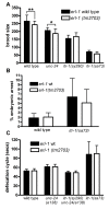
erl-1(tm2703) has no major effect on phenotype of itr-1 mutants. The effect of erl-1(tm2703) on brood size (A), embryonic arrest (B) and defecation cycle length (C) on wild type, unc-24(e138), itr-1(sy290) unc-24(e138) and/or itr-1(sa73) was measured. sy290 is a gain-of-function and sa73 is a weak loss-of-function allele of itr-1. Because itr-1(sy290) is closely linked to unc-24(e138), the phenotype of itr-1(sy290) unc-24(e138) strains was compared to that of strains carrying unc-24(e138) alone. Black bars indicate erl-1 wild type and white bars indicate erl-1(tm2703) genotype. (A) Brood size was determined by counting the number of viable offspring per worm (n = 18-20; * indicates p-value < 0.05; ** indicates p-value < 0.01, one-way ANOVA and Newman-Keuls multiple comparison test). (B) Percentage of offspring arresting as embryos was determined. (C) Defecation cycle length was determined by measuring times between posterior body contractions (pBocs). Values represent the average (+/-SD) of six defecation cycles for each of ten worms (five worms for itr-1(sa73) strains).
Figure 8
Figure 8
erl-1(tm2703) does not alter response to ER-stress. (A) Wild type (wt) or erl-1(tm2703) embryos were plated onto NGM plates containing the indicated concentrations of tunicamycin (TN). After 72 hrs, animals were grouped into three categories (dead, < L4 and ≥ L4). Total number of animals scored are indicated above columns. (B) Mostly adult worms of hsp-4::GFP expressing strains (either erl-1 wt or erl-1(tm2703)) were plated onto NGM plates containing 5 μg/ml TN or DMSO only. Expression of hsp-4::GFP was determined by Western blotting using a GFP specific antibody. Western blots were also probed for ERL-1, demonstrating that ERL-1 protein levels are not altered by ER stress treatment (actin used as loading control).

Similar articles

Cited by

References

    1. Browman DT, Resek ME, Zajchowski LD, Robbins SM. Erlin-1 and erlin-2 are novel members of the prohibitin family of proteins that define lipid-raft-like domains of the ER. J Cell Sci. 2006;119:3149–3160. doi: 10.1242/jcs.03060. - DOI - PubMed
    1. Browman DT, Hoegg MB, Robbins SM. The SPFH domain-containing proteins: more than lipid raft markers. Trends Cell Biol. 2007;17:394–402. doi: 10.1016/j.tcb.2007.06.005. - DOI - PubMed
    1. Pearce MM, Wang Y, Kelley GG, Wojcikiewicz RJ. SPFH2 mediates the endoplasmic reticulum-associated degradation of inositol 1,4,5-trisphosphate receptors and other substrates in mammalian cells. J Biol Chem. 2007;282:20104–20115. doi: 10.1074/jbc.M701862200. - DOI - PubMed
    1. Hoegg MB, Browman DT, Resek ME, Robbins SM. Distinct regions within the erlins are required for oligomerization and association with high molecular weight complexes. J Biol Chem. 2009;284:7766–7776. doi: 10.1074/jbc.M809127200. - DOI - PMC - PubMed
    1. Pearce MM, Wormer DB, Wilkens S, Wojcikiewicz RJ. An endoplasmic reticulum (ER) membrane complex composed of SPFH1 and SPFH2 mediates the ER-associated degradation of inositol 1,4,5-trisphosphate receptors. J Biol Chem. 2009;284:10433–10445. doi: 10.1074/jbc.M809801200. - DOI - PMC - PubMed

Publication types

MeSH terms

Substances

LinkOut - more resources