. 2012 Jan 23:12:5.
doi: 10.1186/1471-213X-12-5.

Regulation of aggregate size and pattern by adenosine and caffeine in cellular slime molds

Affiliations

Regulation of aggregate size and pattern by adenosine and caffeine in cellular slime molds

Pundrik Jaiswal et al. BMC Dev Biol. .

Abstract

Background: Multicellularity in cellular slime molds is achieved by aggregation of several hundreds to thousands of cells. In the model slime mold Dictyostelium discoideum, adenosine is known to increase the aggregate size and its antagonist caffeine reduces the aggregate size. However, it is not clear if the actions of adenosine and caffeine are evolutionarily conserved among other slime molds known to use structurally unrelated chemoattractants. We have examined how the known factors affecting aggregate size are modulated by adenosine and caffeine.

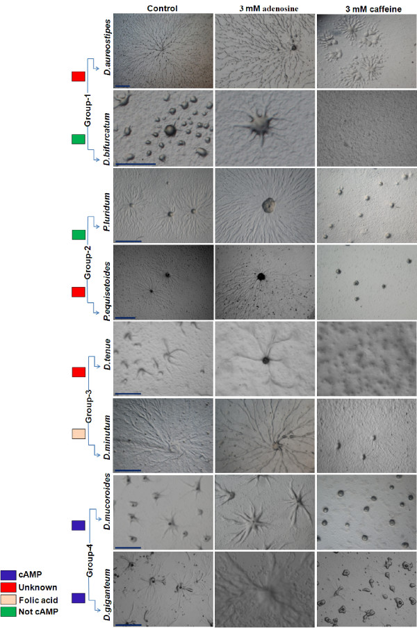
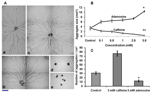
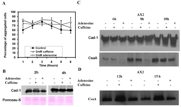
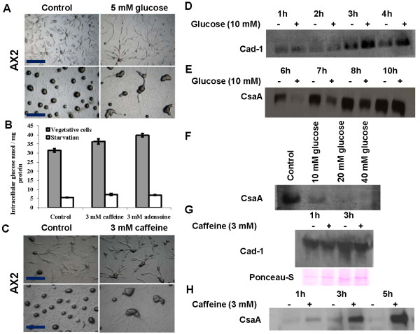
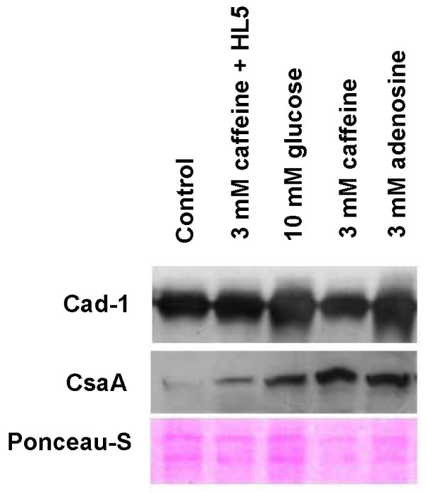
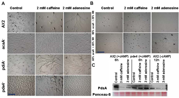
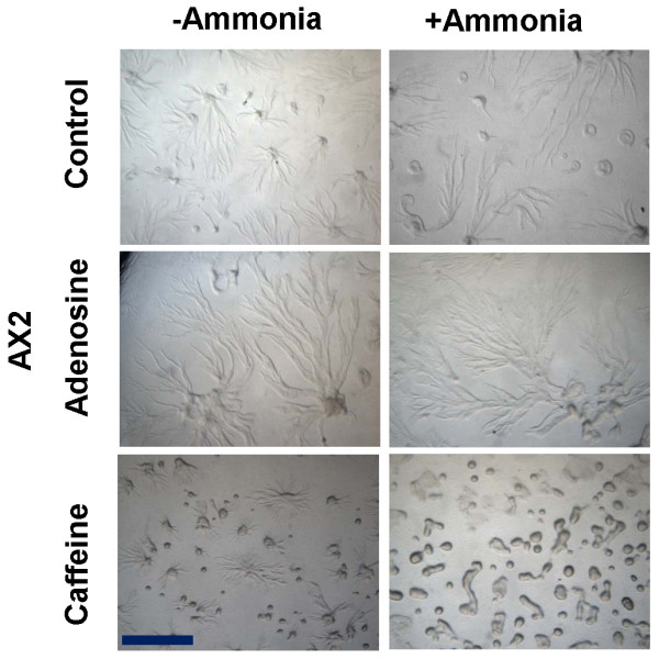
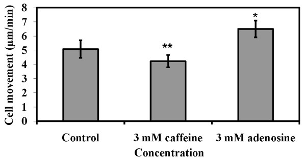
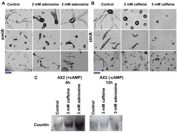
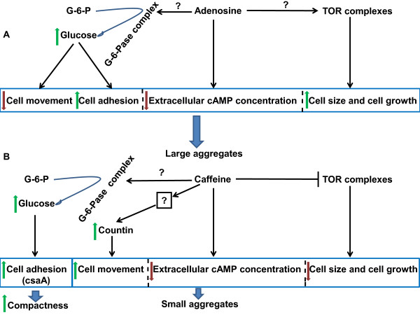
Result: Adenosine and caffeine induced the formation of large and small aggregates respectively, in evolutionarily distinct slime molds known to use diverse chemoattractants for their aggregation. Due to its genetic tractability, we chose D. discoideum to further investigate the factors affecting aggregate size. The changes in aggregate size are caused by the effect of the compounds on several parameters such as cell number and size, cell-cell adhesion, cAMP signal relay and cell counting mechanisms. While some of the effects of these two compounds are opposite to each other, interestingly, both compounds increase the intracellular glucose level and strengthen cell-cell adhesion. These compounds also inhibit the synthesis of cAMP phosphodiesterase (PdsA), weakening the relay of extracellular cAMP signal. Adenosine as well as caffeine rescue mutants impaired in stream formation (pde4- and pdiA-) and colony size (smlA- and ctnA-) and restore their parental aggregate size.

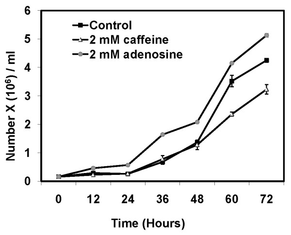
Conclusion: Adenosine increased the cell division timings thereby making large number of cells available for aggregation and also it marginally increased the cell size contributing to large aggregate size. Reduced cell division rates and decreased cell size in the presence of caffeine makes the aggregates smaller than controls. Both the compounds altered the speed of the chemotactic amoebae causing a variation in aggregate size. Our data strongly suggests that cytosolic glucose and extracellular cAMP levels are the other major determinants regulating aggregate size and pattern. Importantly, the aggregation process is conserved among different lineages of cellular slime molds despite using unrelated signalling molecules for aggregation.


Figures

Figure 1
Figure 1
Effect of adenosine and caffeine on the aggregate size of different species of Dictyostelium and Polysphondylium. In all the species examined, large aggregates were formed in the presence of adenosine and contrastingly, with caffeine many small aggregates were formed. The aggregates were developed as described in material and methods. Scale bar = 1000 μm.
Figure 2
Figure 2
Effect of adenosine and caffeine on aggregation pattern of Dictyostelium and Polysphondylium. A) Aggregation pattern of the Polysphondylium cells a) Control (b) with 0.5 mM (c) with 1 mM adenosine. Small aggregate formation in the presence of caffeine (d) 1.0 mM (e) 2.5 mM. Scale bar = 200 μm. Polysphondylium amobae were plated at a density of 1 × 10⁶ cells/cm2 on KK2 plates containing caffeine or adenosine and were allowed to form aggregates in the dark and humid conditions. (B) Aggregate size (Student's t-test, *p < 0.001, **p < 0.004). (C) The number of aggregates (Student's t-test, *p < 0.001). There were few aggregation centres, in the presence of adenosine. Many, small aggregates were formed in the presence of caffeine. Numbers of aggregates were counted in 1 cm2 area. All values represent the mean ± standard error (n = 10).
Figure 3
Figure 3
Cell division kinetics of AX2 cells in presence of 2 mM caffeine or 2 mM adenosine: 2 × 10⁵ cells/ml were inoculated in HL 5 medium containing either adenosine or caffeine with constant shaking at 180 RPM and cell division was estimated by counting the number of cells with a haemocytometer at regular intervals. The values represent mean ± standard deviation (n = 4) (Student's t-test, *p < 0.01, **p < 0.001).
Figure 4
Figure 4
Cell adhesion in Dictyostelium amobae. A) Cell-cell adhesion in the presence of 2 mM adenosine or 2 mM caffeine. It was carried out by scoring for the presence of single cells or clumps of cells with two or more cells adhered to each other. All values represent the mean ± standard error of 7 independent experiments. B) Expression pattern of Cad-1 proteins in AX2 cells. The Cad-1 expression level was checked in cells developed for 2 h and 4 h in Sorensen buffer in the presence or absence of either caffeine or adenosine. C) Expression levels of Cad-1 and CsaA in AX2 cells developed for 6 h, 8 h, 10 h, 12 h and 15 h in the presence or absence of either caffeine or adenosine.
Figure 5
Figure 5
Cells grown in the presence of caffeine led to an increase in the levels of intracellular glucose or developing cells with extracellular glucose led to altered aggregate size and cellular adhesion protein Cad-1 and CsaA expressions: A) Effect of extracellular glucose on aggregate size. B) Cytosolic glucose levels of vegetative and developing cells in the presence or absence of either caffeine or adenosine. The glucose level was estimated as described in materials and methods. The values represent mean ± standard deviation (n = 8), *P < 0.1, **P < 0.01 (Student's t-test). C) Aggregates formed from cells grown in the HL5 medium in the presence or absence of caffeine for the 24 hours and subsequently harvested, washed and developed in the non nutrient agar in the absence of caffeine. D) Cad-1 expression levels in AX2 cells developed for 1 h, 2 h, 3 h and 4 h in Sorensen buffer in the presence or absence of 10 mM glucose. E) CsaA expression levels in cells developed for 6 h, 7 h, 8 h and 10 h in Sorensen buffer in the presence or absence of 10 mM glucose. F) CsaA expression levels in cells developed for 6 h in Sorensen buffer in the presence of different concentrations of glucose. G) Expression pattern of Cad-1 proteins in AX2 cells. Cells were grown in the presence or absence of 3 mM caffeine for 24 hours and Cad-1 expression levels were checked in cells developed for 1 h and 3 h in Sorensen buffer in the absence of caffeine. H) CsaA levels in AX2 cells grown in the presence or absence of caffeine. CsaA levels were checked in the cells developed for 1 h, 3 h and 5 h in Sorensen buffer in the absence of caffeine.
Figure 6
Figure 6
Expression levels of cell adhesion proteins Cad-1 and CsaA during aggregation: The aggregates were developed on non-nutrient agar plates in the presence or absence of caffeine, adenosine or 10 mM glucose. The fully streamed aggregates were washed off with ice chilled Sorenson's buffer and subsequently cells were treated with lysis buffer. 5 μg of the protein was loaded in each well and the equal loading of checked by staining the membrane with Ponceau-S dye. 2nd lane of the blot represents the expression of Cad-1 and CsaA form the aggregate developed in the absence of compounds of the AX2 cells grown in the presence of 3 mM caffeine. The expression levels of these proteins were quantified using image J software (NIH-USA) and the changes in expressions were normalized with loading control.
Figure 7
Figure 7
Caffeine alters cAMP signal relay by affecting cAMP phosphodiesterase synthesis (PdsA). A) Aggregation pattern of AX2, acaA- , pdiA- and pde4- cells in the presence or absence of either caffeine or adenosine. Caffeine rescued the pdiA- and pde4- cells into wild type pheonotype. B) Rescuing of acaA- null phenotype by mixing the AX2 cells with acaA- cells in 4: 1 and 3: 2 ratio and checking the aggregation in presence of either caffeine or adenosine. C) Western blot of PdsA protein, to check the expression levels in vegetative AX2 cells pulsed or unpulsed with cAMP and pde4- cells pulsed with cAMP.
Figure 8
Figure 8
The aggregate formed in the presence of ammonia were comparatively larger than the respective controls. Ammonia was generated by mixing 2 ml of 2 mM NH4Cl solution and 2 ml of 1N NaOH solution [38]. A total of 4 ml solution was poured on the top lid and the dishes having the cells (plated on agar media) were kept inverted. To prevent the diffusion of gas, the plates were tightly sealed with the parafilm. Scale bar = 1000 μm.
Figure 9
Figure 9
Cell movement of AX2 cells in the presence of 2 mM caffeine or 2 mM adenosine. Cell movement assay was performed as described in materials and methods. The values represent mean ± standard deviation from 54 cells obtained from three independent experiments. The significance of experiment was checked performing Student's t-test (*P < 0.03, **P < 0.05).
Figure 10
Figure 10
A) Effect of adenosine on smlA- cells: Adenosine prevented the breaking of the aggregation stream leading to formation of big sized slugs and fruiting bodies. B) Effect of caffeine on countin (ctnA-) null cells: With increasing concentration of caffeine, the size of the aggregate, slug, and fruiting body gets reduced. Scale bar = 200 μm. C) Western blots of Countin in the presence or absence of either caffeine or adenosine.
Figure 11
Figure 11
Model of aggregate size regulation in Dictyostelium by adenosine and caffeine. A) At the aggregation stage, adenosine may be decreasing extracellular cAMP accumulation, favouring cell growth and this might be due to enhanced TOR activity and increased cell movement and cell adhesion by increasing the cytosolic glucose concentration. Collectively all these parameters cause large aggregate formation in the presence of adenosine. B) Inactivation of TOR complex by caffeine leads to reduction in cell number and cell size. Caffeine increases countin expression acting on unknown upstream targets resulting in increased cell movement. Low levels of extracellar cAMP, faster cell movement and few cells available for aggregation are plausible reasons for small sized aggregate formation with caffeine. The compactness of the aggregate is maintained by expression of cell adhesion protein CsaA mainly.

Similar articles

Cited by

References

    1. Bonner JT. The cellular slime molds. Second. Princeton University Press; 1967.
    1. Bonner JT, Barkley DS, Hall EM, Konijn TM, Mason JW, O'Keefe G, Wolfe PB. Acrasin, Acrasinase, and the sensitivity to acrasin in Dictyostelium discoideum. Dev Biol. 1969;20:72–87. doi: 10.1016/0012-1606(69)90005-0. - DOI - PubMed
    1. De Wit RJ, Konijn TM. Identification of the acrasin of Dictyostelium minutum as a derivative of folic acid. Cell Differ. 1983;12:105–210. - PubMed
    1. De Wit RJ, Van Der Velden RJ, Konijn TM. Characterization of the Folic Acid C9-N1o-Cleaving Enzyme of Dictyostelium minutum V3. J Bacteriol. 1983;154:859–863. - PMC - PubMed
    1. Konijn TM, Barkley DS, Chang YY, Bonner JT. Cyclic AMP: a naturally occurring acrasin in the cellular slime molds. Am Nat. 1968;102:225–233. doi: 10.1086/282539. - DOI

Publication types

MeSH terms