. 2012 Jan 23:12:9.
doi: 10.1186/1471-230X-12-9.

Luteolin decreases IGF-II production and downregulates insulin-like growth factor-I receptor signaling in HT-29 human colon cancer cells

Affiliations

Luteolin decreases IGF-II production and downregulates insulin-like growth factor-I receptor signaling in HT-29 human colon cancer cells

Do Young Lim et al. BMC Gastroenterol. .

Abstract

Background: Luteolin is a 3',4',5,7-tetrahydroxyflavone found in various fruits and vegetables. We have shown previously that luteolin reduces HT-29 cell growth by inducing apoptosis and cell cycle arrest. The objective of this study was to examine whether luteolin downregulates the insulin-like growth factor-I receptor (IGF-IR) signaling pathway in HT-29 cells.

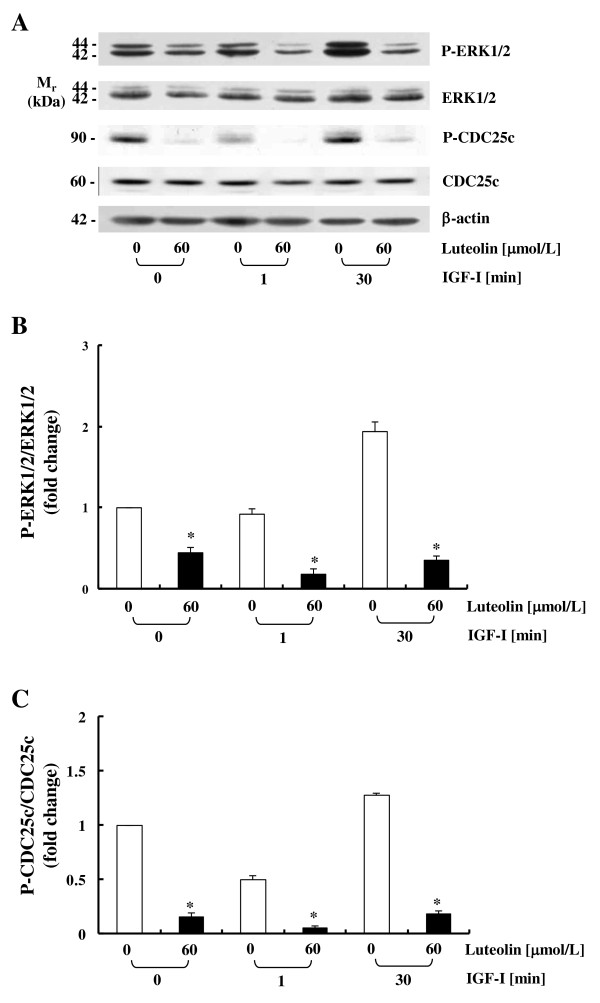
Methods: In order to assess the effects of luteolin and/or IGF-I on the IGF-IR signaling pathway, cells were cultured with or without 60 μmol/L luteolin and/or 10 nmol/L IGF-I. Cell proliferation, DNA synthesis, and IGF-IR mRNA levels were evaluated by a cell viability assay, [3H]thymidine incorporation assays, and real-time polymerase chain reaction, respectively. Western blot analyses, immunoprecipitation, and in vitro kinase assays were conducted to evaluate the secretion of IGF-II, the protein expression and activation of IGF-IR, and the association of the p85 subunit of phophatidylinositol-3 kinase (PI3K) with IGF-IR, the phosphorylation of Akt and extracellular signal-regulated kinase (ERK)1/2, and cell division cycle 25c (CDC25c), and PI3K activity.

Results: Luteolin (0 - 60 μmol/L) dose-dependently reduced the IGF-II secretion of HT-29 cells. IGF-I stimulated HT-29 cell growth but did not abrogate luteolin-induced growth inhibition. Luteolin reduced the levels of the IGF-IR precursor protein and IGF-IR transcripts. Luteolin reduced the IGF-I-induced tyrosine phosphorylation of IGF-IR and the association of p85 with IGF-IR. Additionally, luteolin inhibited the activity of PI3K activity as well as the phosphorylation of Akt, ERK1/2, and CDC25c in the presence and absence of IGF-I stimulation.

Conclusions: The present results demonstrate that luteolin downregulates the activation of the PI3K/Akt and ERK1/2 pathways via a reduction in IGF-IR signaling in HT-29 cells; this may be one of the mechanisms responsible for the observed luteolin-induced apoptosis and cell cycle arrest.


Figures

Figure 1
Figure 1
Luteolin reduces IGF-II secretion dose-dependently in HT-29 human colon cancer cells. (A) HT-29 cells were plated at a density of 2 × 10⁶ cells/100 mm dish in DMEM/F12 supplemented with 10% FBS. After 24 h, cells were serum-starved with serum-free DMEM/F12 supplemented with 5 mg/L of transferrin and 5 μg/L of selenium for 24 h. Cells were treated with various concentrations (0 - 60 μmol/L) of luteolin. 24 h after luteolin treatment, conditioned media were collected and concentrated for immunoblot analysis with an anti-IGF-II antibody. The volumes of media loaded onto the gels were adjusted for equivalent cell numbers. Photographs of the chemiluminescent detection of the blot, which were representative of three independent experiments, are shown. (B) The relative abundance of each band was quantified via densitometric scanning of the exposed films, and the control levels were set at 100%. Each bar represents the mean ± SEM (n = 3). Means without a common letter differ, P < 0.05.
Figure 2
Figure 2
Luteolin abrogates the growth-stimulatory effects of exogenous IGF-I in HT-29 cells. (A) HT-29 cells were plated in 24-well plates at a density of 5 × 10⁴ cells/well. One day later, the cells were serum-starved for 24 h with serum-free DMEM/F12 supplemented with 5 mg/L transferrin, 0.1 g/L BSA, and 5 μg/L selenium for 24 h. After serum-starvation, the cells were incubated in serum-free medium containing 0 or 60 μmol/L of luteolin with or without 10 nmol/L of IGF-I for 24, 48, and 72 h. Viable cell numbers were estimated via an MTT assay. (B) Cells were plated in 96-well plates at a density of 6 × 10³ cells/well. Cells were serum-starved and then treated with 0 or 60 μmol/L of luteolin with or without 10 nmol/L of IGF-I for 2 h. [3H]Thymidine was then added, and the incubation was continued for an additional 1 h in order to measure its incorporation into DNA. Each bar represents the mean ± SEM (n = 6). Means without a common letter differ, P < 0.05.
Figure 3
Figure 3
Luteolin reduces the levels of the IGF-IR protein and mRNA in HT-29 cells. (A) HT-29 cells were plated and treated with luteolin as described in Figure 1 for 2 h. Total cell lysates were prepared and immunoblot analyses were conducted. Photographs of the chemiluminescent detection of the blots, which were representative of three independent experiments, were shown. The relative abundance of IGF-IR to their own β-actin was quantified via densitometric scanning of the exposed films, and the control levels were set at 100%. (B)HT-29 cells were plated and treated with luteolin as described in Figure 1 for 2 h. (C)HT-29 cells were treated with 0 or 60 μmol/L of luteolin for 2, 8, and 24 h. (B, C) Total RNA was isolated and real-time PCR was conducted. Each bar represents mean ± SEM (n = 3). (A, B) Means without a common letter differ, P < 0.05. (C) *Different from 0 μmol/L of luteolin at each treatment time, P < 0.05.
Figure 4
Figure 4
Effects of luteolin on IGF-I-induced tyrosine phosphorylation of IGF-IR, the association of p85 with IGF-IR, and PI3K activity in human colon cancer cells. Cells were plated and cultured as described in Figure 1. (A) HT-29 cells were treated for 2 h with 0 or 60 μmol/L of luteolin and lysed with or without stimulation of 10 nmol/L IGF-I for 0, 1, or 30 minutes. Total cell lysates were incubated with anti-IGF-IRβ antibody and the immune complexes were precipitated with protein A-Sepharose. The immunoprecipitated proteins were analyzed via Western blotting with antibodies raised against phosphotyrosine (PY20), IGF-IRβ, or p85. (B) HT-29 and Caco-2 cells were plated and treated as described above. Total cell lysates were analyzed via Western blotting with an antibody raised against P-IGF-IR. Photographs of the chemiluminescent detection of the blots, which were representative of three independent experiments, were shown. (C) The immune complexes obtained from HT-29 cells were incubated with phosphatidylinositol and [γ-32P]ATP. (D) Active PI3K and luteolin were incubated with phosphatidylinositol and [γ-32P]ATP as described in the Materials and Methods section. Phosphatidylinositol 3-phosphate (PIP) generated by immunoprecipitated PI3K (C) or active PI3Kα (D) was separated via thin-layer chromatography (TLC). An autoradiograph of the TLC plate, which was representative of three independent experiments, is shown. (A, B, C) The relative abundance of each blot was quantified via densitometric scanning of the exposed films and the control levels (0 μmol/L luteolin, without IGF-I stimulation) were set at 100%. Each bar represents the mean ± SEM (n = 3). *Different from 0 μmol/L of luteolin at a stimulation time, P < 0.05.
Figure 5
Figure 5
Luteolin inhibits IGF-I-induced Akt activation in HT-29 cells. Cells were plated and treated as described in Figure 4. Total lysates were prepared and immunoblot analyses were conducted with antibodies raised against p-Akt, Akt, or β-actin. (A) Photographs of the chemiluminescent detection of the blots, which were representative of three independent experiments, were shown. (B) The levels of P-Akt to its own Akt control band on immunoblots were quantified via densitometric scanning of the exposed films, and the control levels (0 μmol/L luteolin, without IGF-I stimulation) were set at 1. Each bar represents the mean ± SEM (n = 3). *Different from 0 μmol/L of luteolin at a stimulation time, P < 0.05.
Figure 6
Figure 6
Luteolin inhibits IGF-I-induced ERK1/2 and CDC25c activation in HT-29 cells. Cells were plated and treated as described in Figure 4. Total lysates were prepared and immunoblot analyses were conducted with antibodies raised against P-ERK1/2, ERK1/2, P-CDC25c, CDC25c, or β-actin. (A) Photographs of the chemiluminescent detection of the blots, which were representative of three independent experiments, were shown. (B, C) The levels of P-ERK1/2 to its own ERK1/2 control band (B) and those of P-CDC25c to its own CDC25c control band (C) on immunoblots were quantified via densitometric scanning of the exposed films, and the control levels (0 μmol/L luteolin, without IGF-I stimulation) were set at 1. Each bar represents the mean ± SEM (n = 3). *Different from 0 μmol/L of luteolin at a stimulation time, P < 0.05.
Figure 7
Figure 7
A tentative scheme for luteolin regulation of the IGF-IR signaling pathway in HT-29 human colon cancer cells. Luteolin reduces the secretion of IGF-II and levels of IGF-IR mRNA and protein, which leads to a reduction in IGF-IR phosphorylation and a subsequent inhibition of PI3K/Akt and ERK1/2/CDC25c activation. Additionally, luteolin directly inhibits PI3K activity. These changes in IGF-I signaling contribute to luteolin-induced apoptosis and cell cycle arrest in HT-29 cells. *From our previously published results [21].

Similar articles

Cited by

References

    1. Roghani M, Lassarre C, Zapf J, Povoa G, Binoux M. Two insulin-like growth factor (IGF)-binding proteins are responsible for the selective affinity for IGF-II of cerebrospinal fluid binding proteins. J Clin Endocrinol Metab. 1991;73(3):658–666. doi: 10.1210/jcem-73-3-658. - DOI - PubMed
    1. Lock K, Pomerleau J, Causer L, Altmann DR, McKee M. The global burden of disease attributable to low consumption of fruit and vegetables: implications for the global strategy on diet. Bull World Health Organ. 2005;83(2):100–108. - PMC - PubMed
    1. Baserga R, Hongo A, Rubini M, Prisco M, Valentinis B. The IGF-I receptor in cell growth, transformation and apoptosis. Biochim Biophys Acta. 1997;1332(3):F105–126. - PubMed
    1. Dupont J, LeRoith D. Insulin and insulin-like growth factor I receptors: similarities and differences in signal transduction. Horm Res. 2001;55(Suppl 2):22–26. - PubMed
    1. Le Roith D, Bondy C, Yakar S, Liu JL, Butler A. The somatomedin hypothesis: 2001. Endocr Rev. 2001;22(1):53–74. doi: 10.1210/er.22.1.53. - DOI - PubMed

Publication types

MeSH terms