

Transfecting the hard-to-transfect lymphoma/leukemia cells using a simple cationic polymer nanocomplex
- PMID: 22269663
- PMCID: PMC3322282
- DOI: 10.1016/j.jconrel.2012.01.007
Transfecting the hard-to-transfect lymphoma/leukemia cells using a simple cationic polymer nanocomplex
Abstract
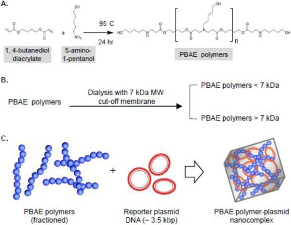
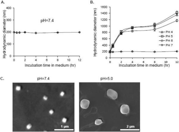
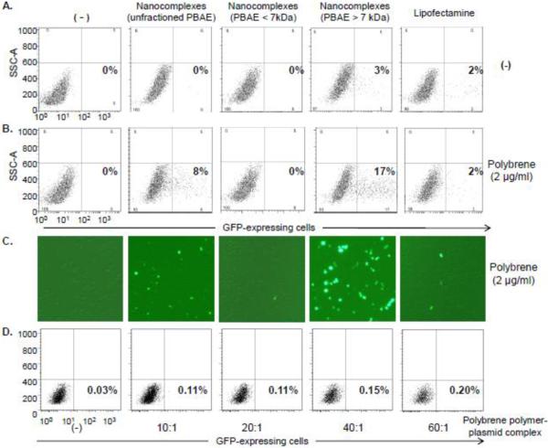
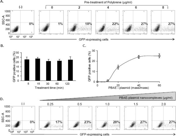
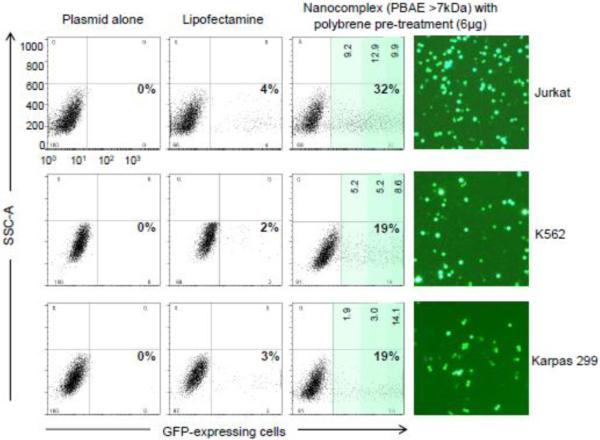
Although the development of gene delivery systems via non-viral-mediated methods is advancing rapidly, it remains a challenge to deliver plasmids into hard-to-transfect cells, such as lymphoma/leukemia cells. To develop an efficient transfection method, we formulated a simple nanocomplex by incorporating poly β-amino ester (PBAE) polymers with plasmid DNAs containing a GFP reporter gene. The formed PBAE-plasmid nanocomplexes are approximately 200nm in diameter and stable under physiological conditions, but become rapidly biodegradable when pH decreases <7.0. Cultured lymphoma/leukemia cells were used for transfection assays and resultant gene delivery rates were determined by quantifying GFP expression. Exposure of cells to the nanocomplexes composed of fractioned PBAE (>7kDa) resulted in GFP expression in 3% of cells, similar to that mediated by the standard Lipofectamine method. However, with polybrene pre-treatment, the nanocomplex could achieve GFP expression in up to 32% of lymphoma/leukemia cells, an 8-fold increase over that mediated by Lipofectamine. These findings demonstrated a simple, efficient method for in vitro gene delivery into hard-to-transfect cells. The nanocomplexes are biodegradable and have minimal cytotoxicity, suggesting the potential use for in vivo gene delivery.
Copyright © 2012 Elsevier B.V. All rights reserved.
Figures

Similar articles
-
Synthesis and application of poly(ethylene glycol)-co-poly(β-amino ester) copolymers for small cell lung cancer gene therapy.Acta Biomater. 2016 Sep 1;41:293-301. doi: 10.1016/j.actbio.2016.05.040. Epub 2016 Jun 1. Acta Biomater. 2016. PMID: 27262740 Free PMC article.
-
Biodegradable brain-penetrating DNA nanocomplexes and their use to treat malignant brain tumors.J Control Release. 2017 Sep 28;262:37-46. doi: 10.1016/j.jconrel.2017.07.009. Epub 2017 Jul 8. J Control Release. 2017. PMID: 28694032 Free PMC article.
-
Continuous microfluidic assembly of biodegradable poly(beta-amino ester)/DNA nanoparticles for enhanced gene delivery.J Biomed Mater Res A. 2017 Jun;105(6):1813-1825. doi: 10.1002/jbm.a.36033. Epub 2017 Apr 12. J Biomed Mater Res A. 2017. PMID: 28177587
-
Development of poly(β-amino ester)-based biodegradable nanoparticles for nonviral delivery of minicircle DNA.ACS Nano. 2013 Aug 27;7(8):7241-50. doi: 10.1021/nn402657d. Epub 2013 Jul 16. ACS Nano. 2013. PMID: 23837668 Free PMC article.
-
Biodegradable polymers as non-viral carriers for plasmid DNA delivery.J Control Release. 2008 Mar 3;126(2):97-110. doi: 10.1016/j.jconrel.2007.10.028. Epub 2007 Dec 4. J Control Release. 2008. PMID: 18201788 Review.
Cited by
-
Megakaryocyte membrane-wrapped nanoparticles for targeted cargo delivery to hematopoietic stem and progenitor cells.Bioeng Transl Med. 2022 Nov 29;8(3):e10456. doi: 10.1002/btm2.10456. eCollection 2023 May. Bioeng Transl Med. 2022. PMID: 37206243 Free PMC article.
-
The challenging nature of primary T lymphocytes for transfection: Effect of protamine sulfate on the transfection efficiency of chemical transfection reagents.Res Pharm Sci. 2020 Oct 19;15(5):437-446. doi: 10.4103/1735-5362.297846. eCollection 2020 Oct. Res Pharm Sci. 2020. PMID: 33628285 Free PMC article.
-
Charge-altering releasable transporters enable phenotypic manipulation of natural killer cells for cancer immunotherapy.Blood Adv. 2020 Sep 8;4(17):4244-4255. doi: 10.1182/bloodadvances.2020002355. Blood Adv. 2020. PMID: 32898247 Free PMC article.
-
Improvement of K562 Cell Line Transduction by FBS Mediated Attachment to the Cell Culture Plate.Biomed Res Int. 2019 Mar 27;2019:9540702. doi: 10.1155/2019/9540702. eCollection 2019. Biomed Res Int. 2019. PMID: 31032368 Free PMC article.
-
Cytosolic Delivery of Macromolecules in Live Human Cells Using the Combined Endosomal Escape Activities of a Small Molecule and Cell Penetrating Peptides.ACS Chem Biol. 2019 Dec 20;14(12):2641-2651. doi: 10.1021/acschembio.9b00585. Epub 2019 Oct 31. ACS Chem Biol. 2019. PMID: 31633910 Free PMC article.
References
-
- Lundstrom K. Latest development in viral vectors for gene therapy. Trends in biotechnology. 2003;21(3):117–122. - PubMed
-
- Woods NB, Muessig A, Schmidt M, Flygare J, Olsson K, Salmon P, Trono D, von Kalle C, Karlsson S. Lentiviral vector transduction of NOD/SCID repopulating cells results in multiple vector integrations per transduced cell: risk of insertional mutagenesis. Blood. 2003;101(4):1284–1289. - PubMed
-
- Wolff JA, Malone RW, Williams P, Chong W, Acsadi G, Jani A, Felgner PL. Direct gene transfer into mouse muscle in vivo. Science (New York, N.Y. 1990;247(4949 Pt 1):1465–1468. - PubMed
-
- Heller LC, Ugen K, Heller R. Electroporation for targeted gene transfer. Expert opinion on drug delivery. 2005;2(2):255–268. - PubMed
Publication types
MeSH terms
Substances
Grants and funding
LinkOut - more resources
Full Text Sources
