. 2012 Mar 16;287(12):8803-15.
doi: 10.1074/jbc.M111.320887. Epub 2012 Jan 23.

Heat shock protein 90α (Hsp90α) is phosphorylated in response to DNA damage and accumulates in repair foci

Affiliations

Heat shock protein 90α (Hsp90α) is phosphorylated in response to DNA damage and accumulates in repair foci

Maria Quanz et al. J Biol Chem. .

Abstract

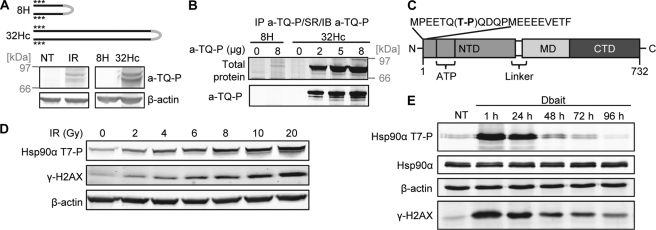
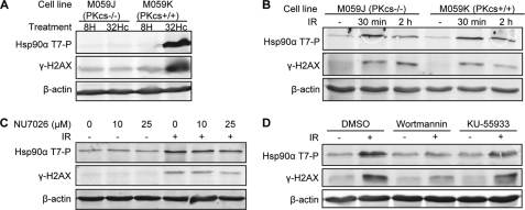
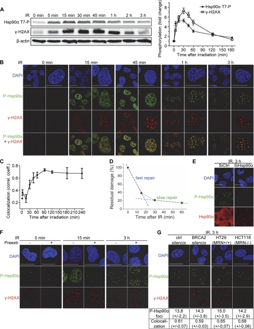
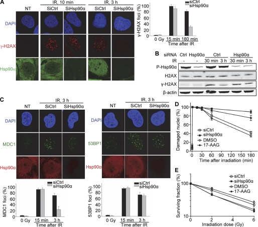
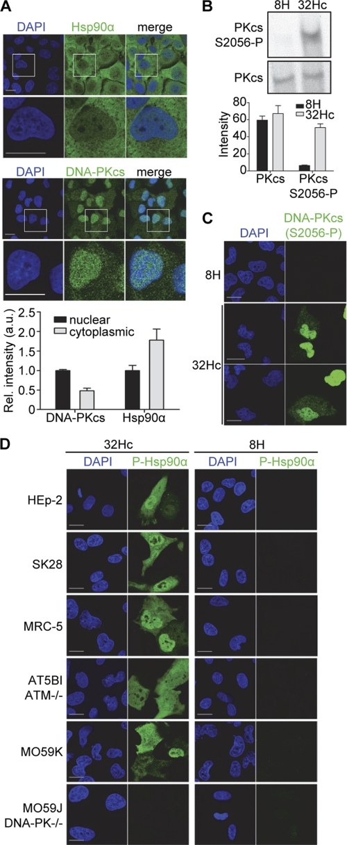
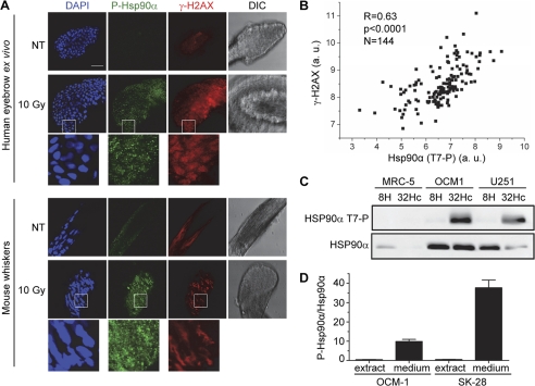
DNA damage triggers a complex signaling cascade involving a multitude of phosphorylation events. We found that the threonine 7 (Thr-7) residue of heat shock protein 90α (Hsp90α) was phosphorylated immediately after DNA damage. The phosphorylated Hsp90α then accumulated at sites of DNA double strand breaks and formed repair foci with slow kinetics, matching the repair kinetics of complex DNA damage. The phosphorylation of Hsp90α was dependent on phosphatidylinositol 3-kinase-like kinases, including the DNA-dependent protein kinase (DNA-PK) in particular. DNA-PK plays an essential role in the repair of DNA double strand breaks by nonhomologous end-joining and in the signaling of DNA damage. It is also present in the cytoplasm of the cell and has been suggested to play a role in cytoplasmic signaling pathways. Using stabilized double-stranded DNA molecules to activate DNA-PK, we showed that an active DNA-PK complex could be assembled in the cytoplasm, resulting in phosphorylation of the cytoplasmic pool of Hsp90α. In vivo, reverse phase protein array data for tumors revealed that basal levels of Thr-7-phosphorylated Hsp90α were correlated with phosphorylated histone H2AX levels. The Thr-7 phosphorylation of the ubiquitously produced and secreted Hsp90α may therefore serve as a surrogate biomarker of DNA damage. These findings shed light on the interplay between central DNA repair enzymes and an essential molecular chaperone.


Figures

FIGURE 1.
FIGURE 1.
Hsp90α phosphorylation at Thr-7 in response to DNA damage. A, top panel, schematic diagram of the structures of the DSB-mimicking 32Hc and the short control 8H. Two complementary DNA strands of 8 bp (8H) or 32 bp (32Hc) are tethered by a hexaethylene glycol linker (gray), and three phosphorothioate nucleotides (asterisks) are incorporated at both the 5′ and 3′ ends to protect the molecule from nucleases. Lower panel, MRC-5 cells were left untreated (NT), irradiated, or transfected with Dbait 32Hc or 8H and lysed 1 h after the end of the treatment. The extracts were immunoblotted and probed with a-TQ-P and β-actin antibodies. B, lysates of MRC-5 cells transfected with Dbait 32Hc or 8H were subjected to immunoprecipitation (IP) with the a-TQ-P antibody. The immunoprecipitates were resolved by SDS-PAGE and stained for total protein with SYPRO Ruby (SR) or immunoblotted (IB) for Hsp90α. C, domain structure of Hsp90α. The phosphorylated Thr-7 in the N terminus of the protein is shown in bold typeface. NTD, N-terminal domain; MD, middle domain; CTD, C-terminal domain; ATP, ATP-binding site. D, MRC-5 cells were irradiated with the indicated doses of γ-irradiation and lysed 30 min after irradiation. The extracts were immunoblotted and probed for Hsp90α (Thr(P)-7), γ-H2AX, and β-actin. E, MRC-5 cells were transfected or not transfected (NT) with Dbait 32Hc and lysed at the indicated time points after transfection. The extracts were treated as in D. Total Hsp90α was detected on a separate membrane.
FIGURE 2.
FIGURE 2.
Involvement of multiple PIKKs in the phosphorylation of Hsp90α at Thr-7. A, M059K and DNA-PK-deficient M059J cells were transfected with Dbait 32Hc or 8H and lysed 1 h after transfection. The extracts were immunoblotted and probed for Hsp90α (Thr(P)-7), γ-H2AX, and β-actin. B, M059K and M059J cells were γ-irradiated (IR) with 10 Gy and lysed at the indicated times after irradiation. The extracts were processed as in A. C, MRC-5 cells were treated 1 h before 10 Gy irradiation (IR) with the indicated concentrations of the DNA-PK inhibitor NU7026. The cells were incubated for 30 min with NU7026 after irradiation and then lysed, and extracts were immunoblotted as in A. D, MRC-5 cells were treated 1 h before irradiation (IR) with 20 μm of the PIKK inhibitor wortmannin, 10 μm of the ATM-inhibitor KU-55933, or vehicle (DMSO). The cells were irradiated (10 Gy), incubated for further 30 min with the inhibitor, and then lysed, and extracts were processed as in A.
FIGURE 3.
FIGURE 3.
Recruitment of phosphorylated Hsp90α to DNA damage sites. A, MRC-5 cells were irradiated (IR) with 10 Gy and lysed at the indicated time points after irradiation. The extracts were subjected to Western blotting, and the blots were probed with antibodies against Hsp90α (Thr(P)-7), γ-H2AX, and β-actin. The graph shows the quantification of H2AX and Hsp90α phosphorylation with standard deviations from three Western blot analyses. B, MRC-5 cells were irradiated with 10 Gy, fixed at the indicated times after IR, and double-immunostained with anti-Hsp90α (Thr(P)-7) (green) and anti-γ-H2AX (red) antibodies. C, colocalization of phosphorylated H2AX and Hsp90α at the indicated times after IR was quantified by measuring Pearson correlation coefficient (correl. coeff.) as described under “Experimental Procedures.” The data are the means ± S.D. >200 cells per time point and two independent experiments. D, cells were irradiated, and DNA damage was quantified at the indicated times by alkaline comet assay. The data shown are the means of median comet tail moments and standard deviations from three independent experiments and are presented as a percentage of initial radiation-induced damage (black squares). Fast and slow repair components are highlighted in blue and green, respectively. E, MRC-5 cells were irradiated with 10 Gy γ-irradiation and fixed at the indicated times after irradiation. Where indicated, cells were extracted with Triton X-100 before fixation. Cells were double-immunostained for Hsp90α (Thr(P)-7) (green) and γ-H2AX (red). F, MRC-5 cells were transfected with 100 nm siRNA against Hsp90α or control siRNA, irradiated 96 h later with 10 Gy γ-irradiation, fixed 3 h after IR, and double-immunostained for Hsp90α (Thr(P)-7) (green) and Hsp90α (red). G, MRN-defective HCT116, MRN-proficient HT29 cells, and HeLa cells silenced with shRNA for BRCA2 (BRCA2 SilenciX) or control shRNA (ctrl SilenciX) were irradiated with 10 Gy, fixed 3 h after IR, and double-immunostained with anti-Hsp90α (Thr(P)-7) (green) and anti-γ-H2AX (red) antibodies. DNA was stained with DAPI (blue). Scale bar, 20 μm. The data show the average foci number with standard deviation as quantified from >200 cells in two independent experiments. Colocalization between Hsp90α (Thr(P)-7) and γ-H2AX was quantified as in C.
FIGURE 4.
FIGURE 4.
Impact of Hsp90α silencing on DNA repair and the organization of repair foci. A–E, MRC-5 cells were transfected with 100 nm siRNA against Hsp90α (SiHsp90α) or equal amounts of control siRNA (SiCtrl) and irradiated with 10 Gy γ-irradiation when indicated. They were then left to grow for 96 h before treatment. A, cells were fixed 3 h after IR (10 Gy) and double-immunostained for γ-H2AX (red) and Hsp90α (green). The bar chart gives the percentage of cells with more than 20 H2AX foci, with a standard deviation for each set of conditions in three independent experiments. B, cells were lysed at the indicated times after IR, and the extracts were immunoblotted and probed for Hsp90α (Thr(P)-7), γ-H2AX, and β-actin. H2AX was detected on a separate membrane. C, cells were fixed 3 h after IR (10 Gy) and double-immunostained for Hsp90α (red) and MDC1 (green) (left panel) or Hsp90α (red) and 53BP1 (green) (right panel). DNA was stained with DAPI (blue). Scale bar, 20 μm. The bar chart gives the percentage of cells with more than 20 foci as in A. D and E, where indicated, cells were treated for 1 h with 250 nm 17-AAG or with vehicle (DMSO) before irradiation. D, cells were irradiated (10 Gy), and damaged nuclei were quantified by the alkaline comet assay at the indicated times. Nuclei were considered damaged when the tail moment exceeded 95% of the values for the nonirradiated control. The data shown are means from three independent experiments with standard deviations. E, cells were γ-irradiated with the indicated doses and quantified 10 days later, as described under “Experimental Procedures.” The data shown are means from three independent experiments with standard deviations.
FIGURE 5.
FIGURE 5.
Cytoplasmic Hsp90α phosphorylation by DNA-PK in response to Dbait 32Hc. A, immunostaining of total Hsp90α and DNA-PK in untreated MRC-5 cells. Data shown are relative mean pixel intensities with standard deviations from cytoplasmic and nuclear regions of >100 cells. The nuclear signal was set to 1. a.u., arbitrary units. B, MRC-5 cells were transfected with Dbait 32Hc or the short inactive 8H and lysed 1 h after the end of the transfection period. The extracts were immunoblotted and probed for DNA-PKcs (S2056-P). DNA-PK was detected on a separate membrane. The bar chart gives quantifications of the signals with standard deviations from three Western blot analyses. C, immunostaining of DNA-PK (S2056-P) (green) in Dbait 32Hc-treated and 8H-treated MRC-5 cells. Cells were fixed 1 h after transfection. D, indicated cell lines were treated with Dbait 32Hc or 8H and immunostained for Hsp90α (Thr(P)-7) (green). DNA was stained with DAPI (blue). Scale bar, 20 μm.
FIGURE 6.
FIGURE 6.
Phosphorylated Hsp90α in tumor xenografts, tissues, and cell supernatants. A, immunostaining of Hsp90α (Thr(P)-7) (green) and γ-H2AX (red) in ex vivo irradiated human eye brow bulbs (upper panel) and whisker bulbs from irradiated mice (lower panel), 1 h after irradiation (10 Gy). DNA was stained with DAPI (blue). Scale bar, 50 μm. DIC, differential interference contrast. a.u., arbitrary units. B, Pearson correlation analysis of Hsp90α (Thr(P)-7) and γ-H2AX levels, as determined from reverse phase protein array analyses of various human tumors (see text) xenografted into mice, as described under “Experimental Procedures.” R and p are Pearson's correlation coefficient and its probability, respectively. C, indicated cells lines were transfected with Dbait 32Hc or 8H and incubated for 24 h without serum. The 50× concentrated cell culture medium was denatured and immunoblotted for Hsp90α (Thr(P)-7). Hsp90α was detected on a separate membrane. D, OCM-1 and SK-28 cells were transfected with Dbait 32Hc and incubated without serum for 24 h. Denatured protein from 50× concentrated cell culture medium and from total cell extracts was immunoblotted and probed for Hsp90α (Thr(P)-7) and Hsp90α. The signals were quantified and the proportion of total Hsp90α that was phosphorylated was calculated.

Similar articles

Cited by

References

    1. Wyman C., Kanaar R. (2006) DNA double strand break repair. All's well that ends well. Annu. Rev. Genet. 40, 363–383 - PubMed
    1. Lieber M. R. (2010) The mechanism of double strand DNA break repair by the nonhomologous DNA end-joining pathway. Annu. Rev. Biochem. 79, 181–211 - PMC - PubMed
    1. Hartlerode A. J., Scully R. (2009) Mechanisms of double strand break repair in somatic mammalian cells. Biochem. J. 423, 157–168 - PMC - PubMed
    1. Pardo B., Gómez-González B., Aguilera A. (2009) DNA repair in mammalian cells. DNA double strand break repair. How to fix a broken relationship. Cell. Mol. Life Sci. 66, 1039–1056 - PMC - PubMed
    1. Ciccia A., Elledge S. J. (2010) The DNA damage response. Making it safe to play with knives. Mol. Cell 40, 179–204 - PMC - PubMed

Publication types

Substances