. 2012 Aug;61(8):1255-68.
doi: 10.1007/s00262-012-1200-1. Epub 2012 Jan 24.

Sensitivity of a novel model of mammary cancer stem cell-like cells to TNF-related death pathways

Affiliations

Sensitivity of a novel model of mammary cancer stem cell-like cells to TNF-related death pathways

Ming Li et al. Cancer Immunol Immunother. 2012 Aug.

Abstract

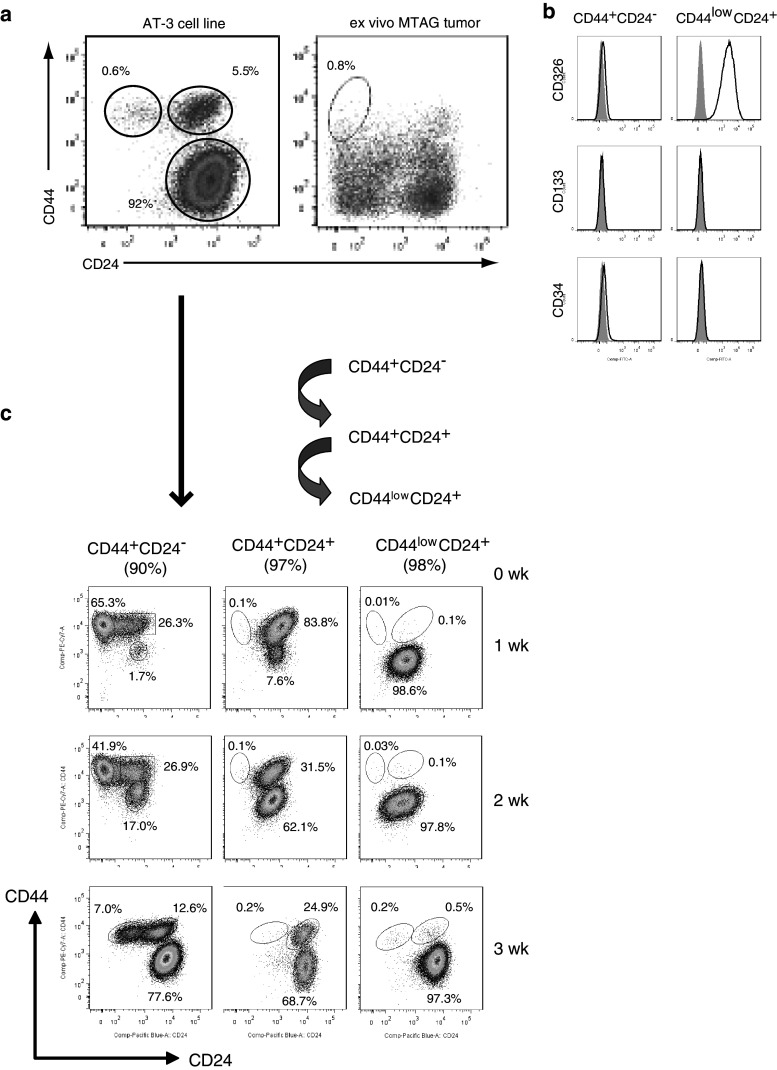
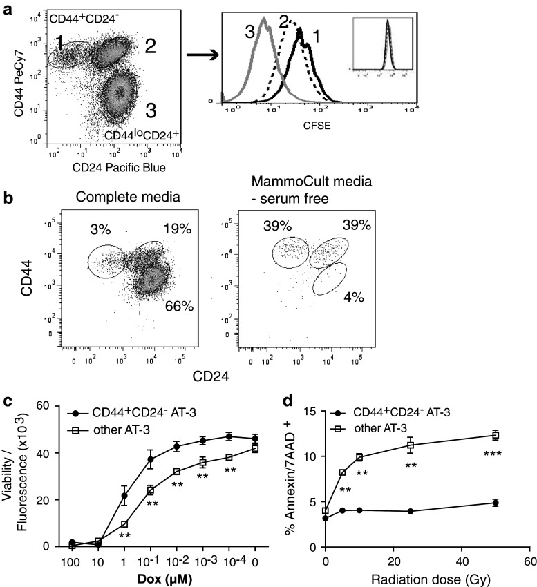
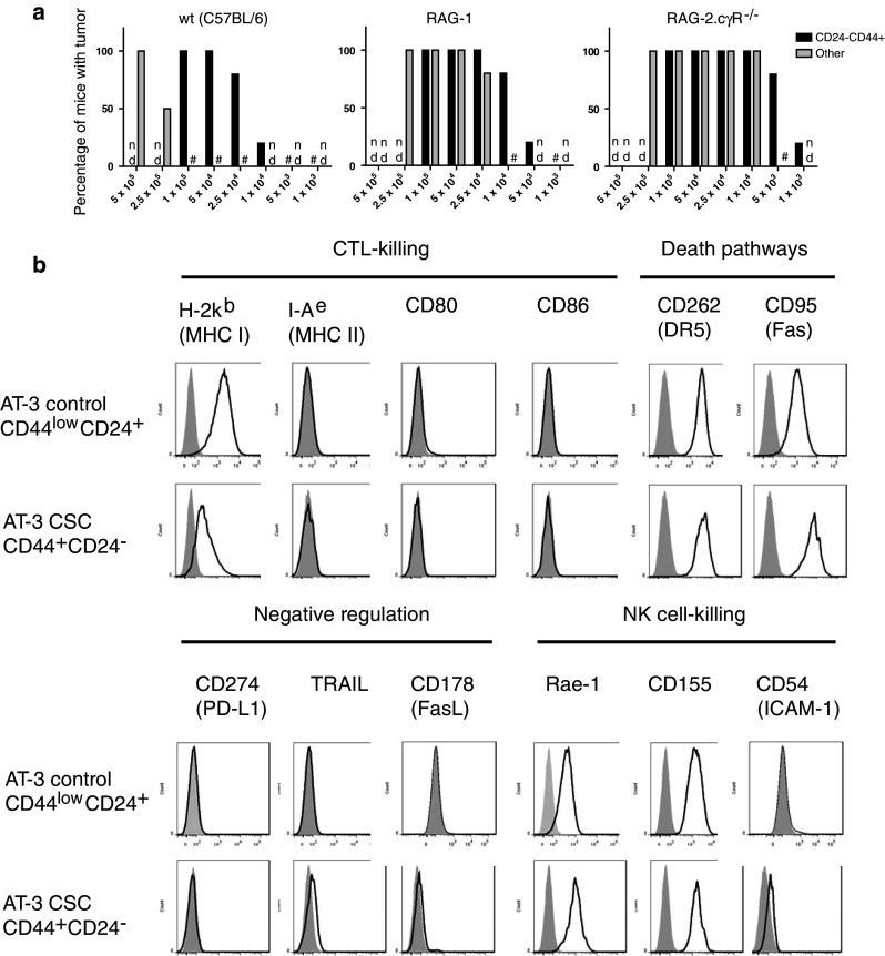
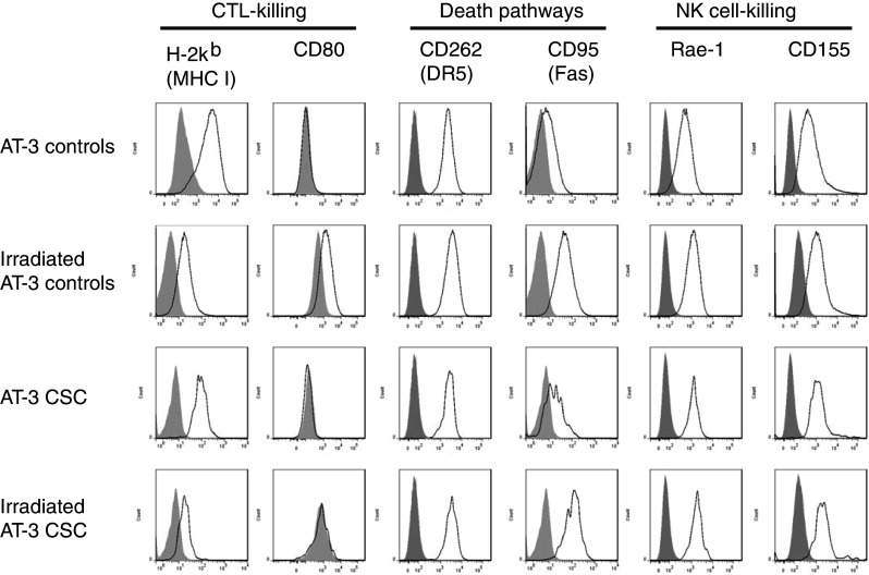
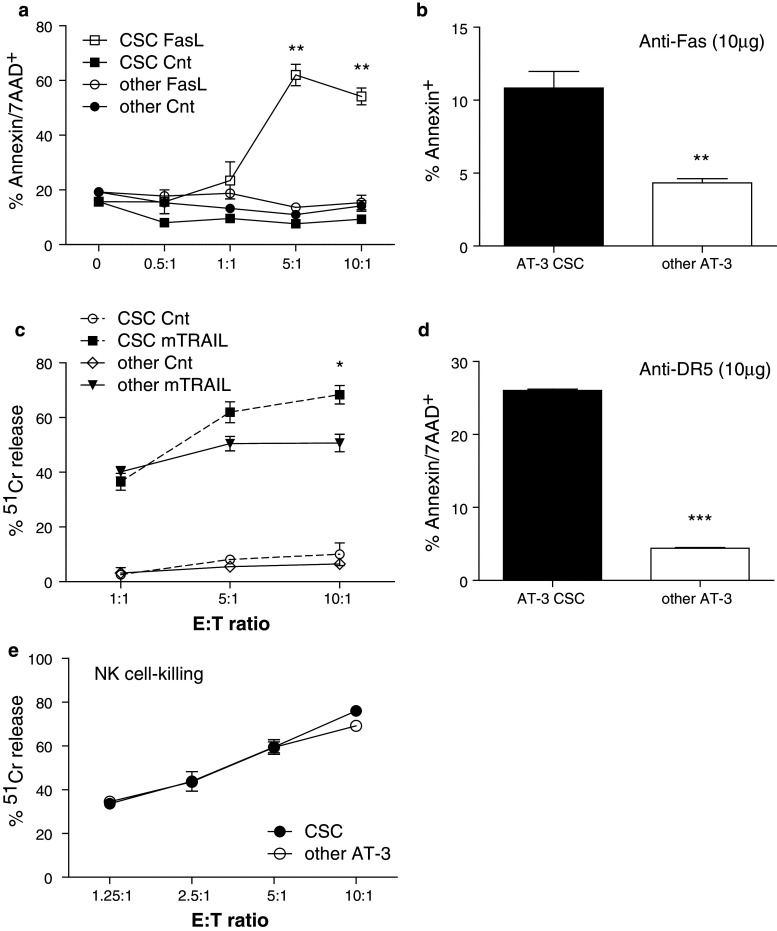
Cancer stem cells (CSC) are resistant to radiation and chemotherapy and play a significant role in cancer recurrence and metastatic disease. It is therefore important to identify alternative strategies, such as immunotherapies that can be used to control this refractory population. A CD44(+)CD24(-/low) subpopulation of cells within the B6 PyMT-MMTV transgenic mouse-derived AT-3 mammary carcinoma cell line was identified, which had CSC-like characteristics, including pluripotency and a resistance to chemo- and radiotherapy. Therefore, unlike xenograph models that require immunocompromised settings, this novel system may provide a means to study immune-mediated responses against CSC-like cells. The immunobiology of the AT-3 CSC-like cell population was studied by their surface molecule expression profile and their sensitivity to specified cell death pathways. Comparable levels of Rae-1, CD155, CD54 and higher levels of Fas and DR5 were expressed on the AT-3 CSC-like cells compared to non-CSC-like tumor cells. Expression correlated with an in vitro sensitivity to cell death by NK cells or through the ligation of the death receptors (Fas or DR5), by their ligands or anti-Fas and anti-DR5 mAbs. Indeed, compared to the rest of the AT-3 tumor cells, the CD44(+)CD24(-/low) subpopulation of cells were more sensitive to both Fas- and TRAIL-mediated cell death pathways. Therefore, despite the refractory nature of CSC to other conventional therapies, these CSC-like cells were not inherently resistant to specified forms of immune-mediated cell death. These results encourage the continued investigation into immunotherapeutic strategies as a means of controlling breast CSC, particularly through their cell death pathways.


Conflict of interest statement

The authors declare that they have no conflict of interest.

Figures

Fig. 1
Fig. 1
a A putative population of CSC-like cells was identified within the AT-3 mammary carcinoma cell line or within single cell preparations of primary MTAG tumors (n > 25) following staining for CD44 and CD24, and flow cytometric analysis. b An analysis of other commonly used cancer stem cell markers, namely CD326, CD133 and CD34 (black line), was conducted on the putative AT-3 CSC-like (CD44+CD24) subpopulation compared to the major CD44CD24+ subpopulation of AT-3 cells. Results are illustrative of at least 4 separate experiments with the gray histogram representing isotype control staining. c The three subpopulations of cells within the AT-3 cell line, based on CD44/CD24 staining, were purified by FACS and cultured under normal AT-3 cell line culturing conditions. The purity of these initial cell preparations is stated in brackets at line 0 week. A sample of each cell culture was taken at weekly intervals over 3 weeks (1–3 weeks) and reassessed for CD44/CD24 expression. Results are representative of 2 separate experiments
Fig. 2
Fig. 2
a An AT-3 cell line that had been enriched for CD44+CD24 cells (approx. 3%) was labeled with CFSE (See histogram inset). After 72-h incubation, cells were harvested and analyzed by flow cytometry to determine CFSE dilution within the three CD44/CD24 cell subpopulations. Data represent one of three similar experiments. b The AT-3 cell line, enriched for CD44+CD24 cells (approx. 5%), was plated in non-adherent plates either in normal DMEM complete media or in serum-free MammoCult® media. After 3-day incubation, the resulting cells were harvested and analyzed for CD44/CD24 expression. Experiment was repeated with similar results. c CD44+CD24 cells were purified by FACS and their sensitivity to Doxorubicin compared to that of the “other” AT-3 cells (i.e., AT-3 minus CD44+CD24 cells). Cells were incubated in triplicate wells, with a dose titration of Doxorubicin for 20 h and the amount of viable cells remaining determined by fluorescence intensity of the CellTiter-Blue® viability assay reagent. Results represent the mean ± SEM of fluorescence readings of two similar experiments. Significance determined by Student’s t test, where ** represents a P value between 0.01 and 0.001. d Similar to (c), cells were purified and irradiated for various exposure times from 5 to 50 Gy. The CD44+CD24 and other AT-3 cell population were then plated and incubated for 20 h before harvesting and Annexin/7-AAD staining. Cells were analyzed by flow cytometry and the percentage of Annexin and 7-AAD double-positive cells determined. Results are representative of two separate experiments, and the P value determined using linear regression
Fig. 3
Fig. 3
a AT-3 cells were sorted into two cell populations based on CD44/CD24 staining and used to challenge wt, RAG-1 and RAG-2.cγR−/− (common-gamma-chain receptor null) mice in the mammary fatpad at a range of cell doses (n = 5–6 mice per group). The “other” population of cells consists of the remaining AT-3 cells minus the CD44+CD24 subpopulation. Mice were monitored for progressive tumor growth. # represents groups that did not develop tumors; nd = not determined. b Sorted AT-3 CD44/CD24 subpopulations were stained for a range of immunologically relevant surface markers and their expression levels determined. Specific marker expression is represented by the black line and isotype staining by the shaded gray area. Results are representative of at least 3 individual experiments
Fig. 4
Fig. 4
The AT-3 cell line was irradiated with 50 Gy, washed and cultured a further 96 h. Cells were harvested and stained for CD44/CD24 and a range of other immunologically relevant surface markers. Cells were then gated on either CD44+CD24 (AT-3 CSC) or CD44−/lowCD24+ (control) populations and their surface marker expression levels determined. Specific marker expression is represented by the black line and isotype staining by the shaded gray area. Results are representative of at least 3 separate experiments
Fig. 5
Fig. 5
a L5178Y-mFasL or control “effectors” were added at stated ratios to AT-3 CSC or other (AT-3 minus CSC) target cells and incubated 20 h. Cells were harvested from wells and stained with Annexin and 7-AAD before analysis by flow cytometry. Graph represents the mean ± SEM of Annexin/7-AAD double-positive cells from duplicate wells and where ** equals a P value of 0.005. Data are representative of three separate killing assays. b Anti-Fas mAb or its isotype control (10 μg/ml) was added to triplicate wells of sorted AT-3 CSC or other AT-3 cells at 5 × 104 cells/well and incubated for 20 h. Cells were harvested and stained for Annexin before flow cytometric analysis. Graph represents the mean ± SEM of Annexin positive cells and where ** equals a P value of 0.0052 calculated using a Student’s t test. Data are representative of three separate experiments. c Standard 4-h chromium release assays were conducted using 2PK3-mTRAIL and control “effector” cells against the AT-3 CSC or other AT-3 target cell populations. Graph represents the mean ± SEM of % lysis from triplicate wells. Data are representative of three separate killing assays and * equals a P value of 0.019 when comparing mTRAIL-induced killing of CSC and other AT-3 cells. d The MD5.1 (anti-DR5) mAb, or its Ig control, was added to P815 cells (FcR+CD16+) at 10 μg/ml and combined with AT-3 CSC or other AT-3 target cell populations at an E:T of 10:1. Following an overnight incubation, cells were harvested and stained for CD16, Annexin and 7AAD. The percentage of Annexin/7AAD double-positive cells within the CD16 target cell populations was determined. The graphed values are the mean ± SEM of duplicate wells that have had the background Ig percentage subtracted. The results are representative of 3 separate MD5.1-induced killing assays. *** equals a P value of <0.0001. e NK effector cells were purified from syngeneic spleens and stimulated in vitro with IL-2 prior to mixing with 51Cr-labeled AT-3 CSC and other AT-3 target cells in a standard 4-h chromium release assay. Graph represents the mean ± SEM of % lysis from triplicate wells at a range of E/T ratios. Supernatants were harvested, the amount of 51Cr release counted and the percentage lysis determined. Results represent at least 3 similar NK killing assays

Similar articles

Cited by

References

    1. Cobaleda C, Cruz JJ, Gonzalez-Sarmiento R, Sanchez-Garcia I, Perez-Losada J. The emerging picture of human breast cancer as a stem cell-based disease. Stem Cell Rev. 2008;4:67–79. doi: 10.1007/s12015-008-9012-6. - DOI - PubMed
    1. Dontu G, Liu S, Wicha MS. Stem cells in mammary development and carcinogenesis: implications for prevention and treatment. Stem Cell Rev. 2005;1:207–213. doi: 10.1385/SCR:1:3:207. - DOI - PubMed
    1. Baumann M, Krause M, Hill R. Exploring the role of cancer stem cells in radioresistance. Nature Rev Cancer. 2008;8:545–554. doi: 10.1038/nrc2419. - DOI - PubMed
    1. Eyler CE, Rich JN. Survival of the fittest: cancer stem cells in therapeutic resistance and angiogenesis. J Clin Oncol Off J Am Soc Clin Oncol. 2008;26:2839–2845. doi: 10.1200/JCO.2007.15.1829. - DOI - PMC - PubMed
    1. Li X, Lewis MT, Huang J, et al. Intrinsic resistance of tumorigenic breast cancer cells to chemotherapy. J Natl Cancer Inst. 2008;100:672–679. doi: 10.1093/jnci/djn123. - DOI - PubMed

Publication types

MeSH terms