

Magnetic resonance imaging of glutamate
- PMID: 22270722
- PMCID: PMC3274604
- DOI: 10.1038/nm.2615
Magnetic resonance imaging of glutamate
Abstract
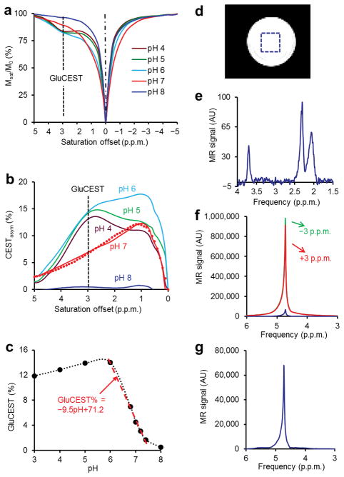
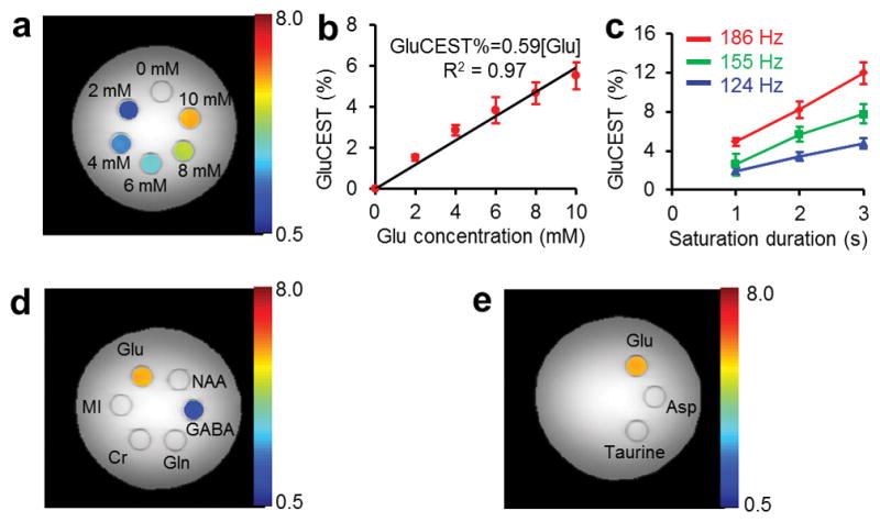
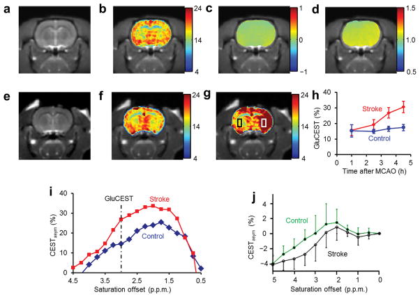
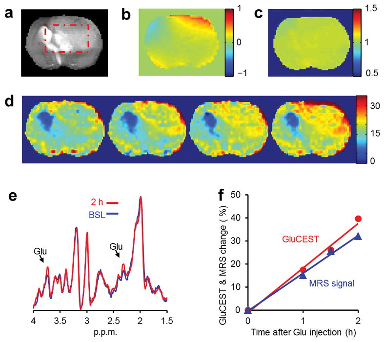
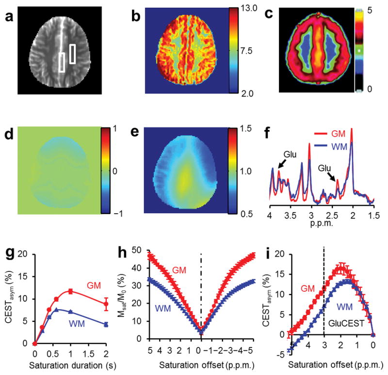
Glutamate, a major neurotransmitter in the brain, shows a pH- and concentration-dependent chemical exchange saturation transfer effect (GluCEST) between its amine group and bulk water, with potential for in vivo imaging by nuclear magnetic resonance. GluCEST asymmetry is observed ∼3 p.p.m. downfield from bulk water. Middle cerebral artery occlusion in the rat brain resulted in an ∼100% elevation of GluCEST in the ipsilateral side compared with the contralateral side, predominantly owing to pH changes. In a rat brain tumor model with blood-brain barrier disruption, intravenous glutamate injection resulted in a clear elevation of GluCEST and a similar increase in the proton magnetic resonance spectroscopy signal of glutamate. GluCEST maps from healthy human brain were also obtained. These results demonstrate the feasibility of using GluCEST for mapping relative changes in glutamate concentration, as well as pH, in vivo. Contributions from other brain metabolites to the GluCEST effect are also discussed.
Figures
References
-
- Petroff OA. GABA and glutamate in the human brain. Neuroscientist. 2002;8:562–573. - PubMed
-
- Harrison PJ. Metabotropic glutamate receptor agonists for schizophrenia. Br J Psychiatry. 2008;192:86–87. - PubMed
-
- Paul IA, Skolnick P. Glutamate and depression: clinical and preclinical studies. Ann N Y Acad Sci. 2003;1003:250–272. - PubMed
-
- Chojnacka-Wojcik E, Klodzinska A, Pilc A. Glutamate receptor ligands as anxiolytics. Curr Opin Investig Drugs. 2001;2:1112–1119. - PubMed
Publication types
MeSH terms
Substances
Grants and funding
LinkOut - more resources
Full Text Sources
Other Literature Sources
Medical
