Review
. 2012 Dec;7(4):820-34.
doi: 10.1007/s11481-011-9336-x. Epub 2012 Jan 21.

CXCL12 signaling in the development of the nervous system

Affiliations
Review

CXCL12 signaling in the development of the nervous system

Divakar S Mithal et al. J Neuroimmune Pharmacol. 2012 Dec.

Abstract

Chemokines are small, secreted proteins that have been shown to be important regulators of leukocyte trafficking and inflammation. All the known effects of chemokines are transduced by action at a family of G protein coupled receptors. Two of these receptors, CCR5 and CXCR4, are also known to be the major cellular receptors for HIV-1. Consideration of the evolution of the chemokine family has demonstrated that the chemokine Stromal cell Derived Factor-1 or SDF1 (CXCL12) and its receptor CXCR4 are the most ancient members of the family and existed in animals prior to the development of a sophisticated immune system. Thus, it appears that the original function of chemokine signaling was in the regulation of stem cell trafficking and development. CXCR4 signaling is important in the development of many tissues including the nervous system. Here we discuss the manner in which CXCR4 signaling can regulate the development of different structures in the central and peripheral nervous systems and the different strategies employed to achieve these effects.


Figures

Fig. 1
Fig. 1
The figure illustrates the distribution of EGFP expression in an E 11 embryo from a CXCR4-EGFP BAC transgenic reporter mouse, showing the wide distribution of CXCR4 expression in the developing nervous system and in other structures (unpublished observations)
Fig. 2
Fig. 2
CXCR4 signaling in the development of the mouse dentate gyrus (DG). Left hand panel: Expression of SDF1 and CXCR4 in developing neocortex and hippocampus. A–G: Coronal sections through mouse forebrains on E15.5, E17.5, and the day of birth (P0) processed for in situ hybridization. Panel G shows a higher magnification of the DG (dg) region. SDF1 is expressed in the intermediate zone (iz) of the neocortex (ncx) (A), in the meninges (mng) overlying the hippocampus (hp) (A, C, E, and G), and in a zone close to the hippocampal fissure also occupied by Cajal–Retzius cells (A, C, E, and G; see text). White arrowhead in G points to the opening of the hippocampal fissure. White asterisk in G marks some of the SDF1-expressing cells lining the hippocampal fissure. CXCR4 also is expressed in the iz at E15.5 (B). At all ages, strongest CXCR4 expression appears in the developing DG and in a migratory stream (ms) of cells moving toward the DG (D and F). Right hand panel: Defects in the secondary proliferative cell population that forms the DG. A-D: Coronal sections through the E18.5 hippocampus of wild-type (A and C) or CXCR4 mutant mice (B and D), processed to show expression of Prox1 (blue) or Prox1 together with BrdUrd-labeled dividing cells (orange). In the wild-type mouse, Prox1 is expressed in the forming DG (A and C). By contrast, in the mutant, Prox1 is expressed in the vestigial DG but also along the migratory stream (ms) (arrows in D) of dividing cells running along the ventral surface of the hippocampus into the DG. BrdUrd-labeled cells of the ms can be seen between the two arrows in C. (E) A higher magnification of the migrating stream of cells shown in D. Numerous blue, Prox1-expressing cells appear among the brown, BrdUrd-labeled cells, but the populations appear largely distinct. Arrows in E point to two single-labeled cells. (F and G) High magnification views of BrdUrd-labeled dividing cells (dark blue) coursing through the ms in a wild-type (F) and a CXCR4 mutant mouse (G). About 30% fewer BrdUrd-labeled cells appear in the ms of the mutant (G) than in the wild type (Lu et al. 2002)
Fig. 3
Fig. 3
Heterogeneity of EGFP-expressing cells in the dentate gyrus (DG) of CXCR4-EGFP BAC transgenic mice during postnatal development. EGFP is expressed in the DG as well as in a population of cells with the position and morphology of Cajal-Retzius cells. At 1 (A) and 2 (B) weeks, CXCR4 is expressed throughout the entire DG, more numerous cells being observed in the inner layer of the granular layer as well as in the SGZ. During development, the concentration of EGFP-expressing cells decreased in the outermost parts of the granule cell layer. At 3–5 weeks (D–I), CXCR4-EGFP-expressing cells are more localized to the SGZ and internal aspects of the granule cell layer. F: The expression pattern of CXCR4-EGFP cells in Cajal-Retzius cells. At 6 weeks (J–M), CXCR4 is expressed mainly in immature granule cells. In addition, it is also expressed in some neural progenitors (type 2 cells, insert in M) and radial astrocyte-like cells (type 1 cells) localized in the SGZ and extending long processes into the granular cell layer, as shown at higher magnification in M. At 3 months (N, O), the expression of CXCR4 is more restricted. Scale bars: 200 μm in A, B; 100 μm in C, D; 50 μm in E, F, G, H, I, J, K, L, N, O; 20 μm in M (Tran et al. 2007)
Fig. 4
Fig. 4
The expression patterns of SDF1, CXCR4, and Sox10 within the spinal cords of mouse embryos. P1, A, B, Coronal sections through the wild-type mouse mid-spinal cord at E9.5, processed with in situ hybridization to show mRNA expression of SDF1 and CXCR4. SDF1 expression extends close to the site at which neural crest cells emerge from the neural tube (A, asterisk). The latter cells are marked by prominent expression of CXCR4 (B, arrowheads). Marked expression also appears in a distinct region of the ventral spinal cord, which may approximately correspond to the position of developing motor neurons. P2, A–F, Migrating DRG neural crest cells are CXCR4 positive. Sections through the spinal cord of a CXCR4 mutant (A–C) and a littermate control (D–F) at E11. Sections were processed for double-fluorescence in situ hybridization (Sox10, green, encodes a transcription factor expressed in neural crest cells; CXCR4, red). A–C, In a CXCR4 homozygous mutant, single fluorescence for Sox10 marks neural crest cells in and migrating to the DRGs on either side of the spinal cord (A). Arrowheads point to a patch of migrating cells also labeled for CXCR4 fluorescence (B, C). C is an overlay of A and B. D–F, Higher magnification of a Sox10- (D, F; arrowhead) and CXCR4-(E, F) expressing cell group migrating toward the DRG (F is an overlay of D, E). P3, A–C, Coronal sections through wild-type E14.5 spinal cord processed with one- or two-color in situ hybridization for SDF1 (blue in A) or both SDF1 (brown in B, C) and CXCR4 (blue in B, C). SDF1 expression is strong over the dorsal neural tube and runs ventrally in two streams that appear to surround the DRGs (B, C; arrowheads). Note that at this later age, E14.5, neural crest migration to the DRGs is near completion (Belmadani et al. 2005)
Fig. 5
Fig. 5
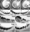
Abnormal DRG development in the absence of CXCR4 receptors. Whole-mount E12 embryos processed to show DRG expression of TrkA (A–C, D, E) and TrkC (F, G). A’–C’ are high-magnification views of the bottom third of A–C. In contrast with the regularly shaped DRGs forming in control embryos (A), DRGs in CXCR4 null mutants are misshapen, disorganized, or small (B, C). Arrowheads indicate normal DRGs (A), compared with split, misshapen DRGs and cell islands in B and C. Ectopic cells dorsal to the DRGs are indicated in E but are absent from the more regular DRGs in a control mouse (D). Similarly, DRGs marked by expression of TrkC are regularly shaped in a control mouse (F) but split or malformed in a CXCR4 null mouse (G) (Belmadani et al. 2005)

Comment in

Similar articles

Cited by

References

    1. Alcendor DJ, Zong J, Dolan A, Gatherer D, Davison AJ, Hayward GS. Patterns of divergence in the vCXCL and vGPCR gene clusters in primate cytomegalovirus genomes. Virology. 2009;395:21–32. - PMC - PubMed
    1. Alonso E, Gomez-Santos L, Madrid JF, Saez F. The expression of a novel cxcr4 gene in xenopus embryo. Histol Histopathol. 2009;24:1097–1103. - PubMed
    1. Ara T, Nakamura Y, Egawa T, Sugiyama T, Abe K, Kishimoto T, Matsui Y, Nagasawa T. Impaired colonization of the gonads by primordial germ cells in mice lacking a chemokine, stromal cell-derived factor-1 (SDF1). Proc Natl Acad Sci U S A. 2003;100:5319–5323. - PMC - PubMed
    1. Bagri A, Gurney T, He X, Zou YR, Littman DR, Tessier-Lavigne M, Pleasure SJ. The chemokine SDF regulates migration of dentate granule cells. Development. 2002;129:4249–4260. - PubMed
    1. Bajetto A, Barbero S, Bonavia R, Piccioli P, Pirani P, Florio T, Schettini G. Stromal cell-derived factor 1 alpha induces astrocyte proliferation through the activation of extracellular signal-regulated kinases 1/2 pathway. J Neurochem. 2001a;77:1226–1236. - PubMed

LinkOut - more resources