

Estrogen receptor-β in the gonadotropin-releasing hormone neuron
- PMID: 22271291
- PMCID: PMC9335859
- DOI: 10.1055/s-0031-1299594
Estrogen receptor-β in the gonadotropin-releasing hormone neuron
Abstract
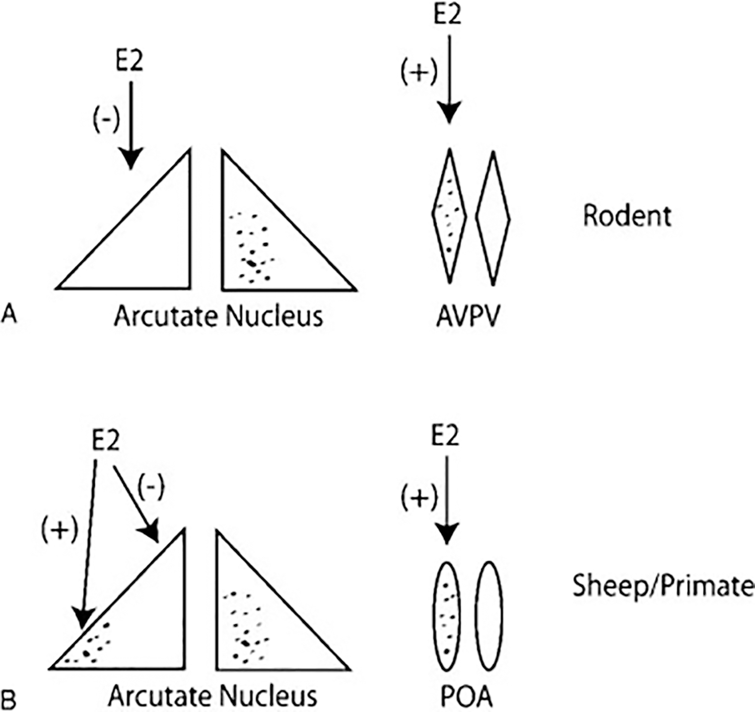
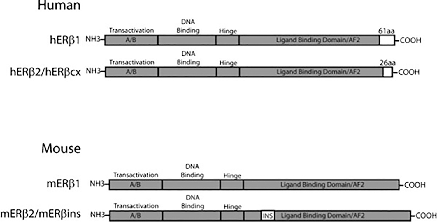
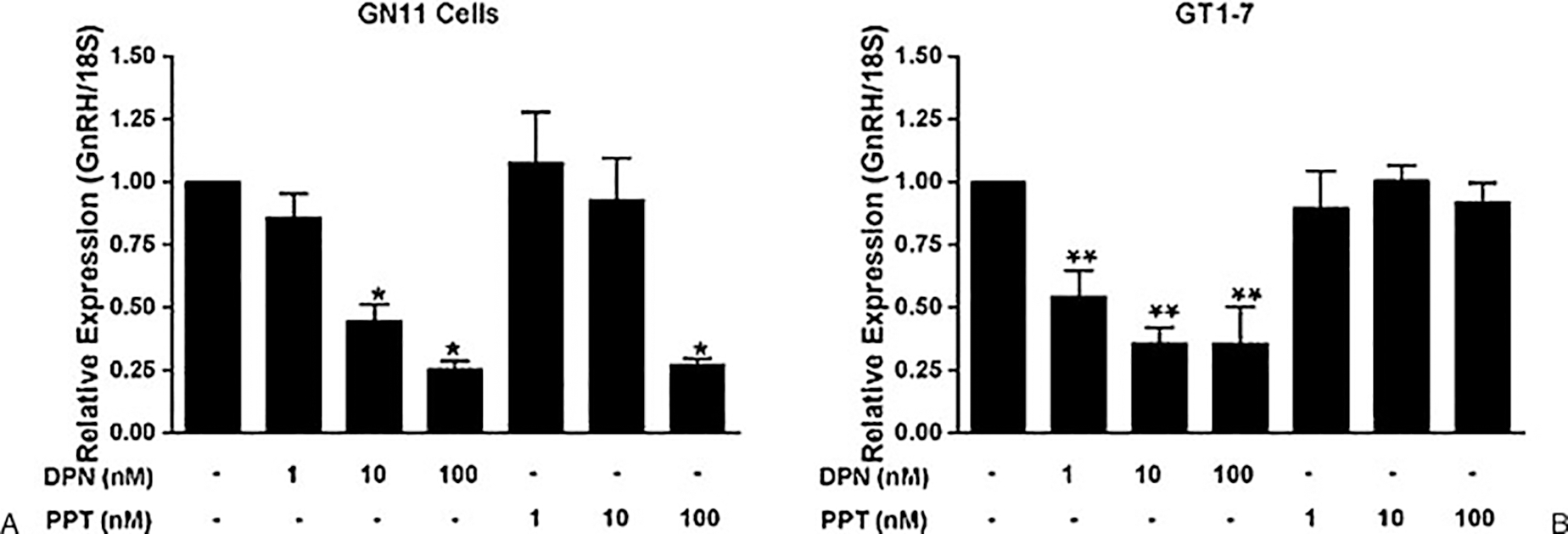
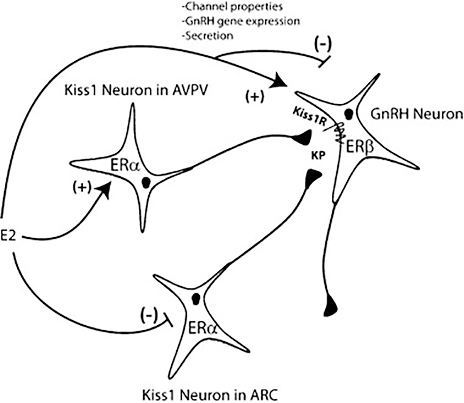
Estrogen regulation of gonadotropin-releasing hormone (GnRH) neuronal activity plays a crucial role in homeostatic regulation of the hypothalamic-pituitary-gonadal axis. Estrogen also coordinates a complex series of physiological changes culminating with a surge of gonadotropin secretion that triggers ovulation of a developed follicle from the ovary. The coordinated functions of estrogen ensure that the female will elaborate appropriate reproductive behaviors ultimately designed to deliver sperm to the oocyte and to provide a receptive uterine environment for the fertilized embryo. Although the effects of estrogen on GnRH neuronal function have long been proposed to be indirect due to the presumed lack of estrogen receptors in GnRH neurons, the identification of alternative estrogen signaling pathways, including estrogen receptor (ER)β and membrane ERs such as GPR30, has put the focus back on estrogen's effect at the level of the GnRH neuron itself. One candidate to mediate the effects of estrogen is the β isoform of the estrogen receptor. We review the evidence for a role for ERβ-mediated regulation of GnRH neuronal function.
Thieme Medical Publishers 333 Seventh Avenue, New York, NY 10001, USA.
Figures
Similar articles
-
Converse regulatory functions of estrogen receptor-alpha and -beta subtypes expressed in hypothalamic gonadotropin-releasing hormone neurons.Mol Endocrinol. 2008 Oct;22(10):2250-9. doi: 10.1210/me.2008-0192. Epub 2008 Aug 13. Mol Endocrinol. 2008. PMID: 18701637 Free PMC article.
-
Definition of estrogen receptor pathway critical for estrogen positive feedback to gonadotropin-releasing hormone neurons and fertility.Neuron. 2006 Oct 19;52(2):271-80. doi: 10.1016/j.neuron.2006.07.023. Neuron. 2006. PMID: 17046690 Free PMC article.
-
Impairments in the reproductive axis of female mice lacking estrogen receptor β in GnRH neurons.Am J Physiol Endocrinol Metab. 2018 Nov 1;315(5):E1019-E1033. doi: 10.1152/ajpendo.00173.2018. Epub 2018 Jul 24. Am J Physiol Endocrinol Metab. 2018. PMID: 30040478 Free PMC article.
-
Ovarian actions of estrogen receptor-β: an update.Semin Reprod Med. 2012 Jan;30(1):32-8. doi: 10.1055/s-0031-1299595. Epub 2012 Jan 23. Semin Reprod Med. 2012. PMID: 22271292 Review.
-
Estrogen Receptors Modulation of Anxiety-Like Behavior.Vitam Horm. 2017;103:27-52. doi: 10.1016/bs.vh.2016.08.004. Epub 2016 Oct 13. Vitam Horm. 2017. PMID: 28061972 Free PMC article. Review.
Cited by
-
Estrogen receptor β exon 3-deleted mouse: The importance of non-ERE pathways in ERβ signaling.Proc Natl Acad Sci U S A. 2015 Apr 21;112(16):5135-40. doi: 10.1073/pnas.1504944112. Epub 2015 Apr 6. Proc Natl Acad Sci U S A. 2015. PMID: 25848008 Free PMC article.
-
Effects of menopause on temperature regulation.Temperature (Austin). 2025 Apr 23;12(2):92-132. doi: 10.1080/23328940.2025.2484499. eCollection 2025. Temperature (Austin). 2025. PMID: 40330614 Free PMC article. Review.
-
Kisspeptin restores pulsatile LH secretion in patients with neurokinin B signaling deficiencies: physiological, pathophysiological and therapeutic implications.Neuroendocrinology. 2013;97(2):193-202. doi: 10.1159/000336376. Epub 2012 Feb 24. Neuroendocrinology. 2013. PMID: 22377698 Free PMC article. Clinical Trial.
-
Exploration of the molecular mechanism of modified Danggui Liuhuang Decoction in treating central precocious puberty and its effects on hypothalamic-pituitary-gonadal axis hormones.Hereditas. 2025 Apr 8;162(1):56. doi: 10.1186/s41065-025-00420-9. Hereditas. 2025. PMID: 40200320 Free PMC article.
-
Assessing hypothalamic pituitary gonadal function in reproductive disorders.Clin Sci (Lond). 2023 Jun 14;137(11):863-879. doi: 10.1042/CS20220146. Clin Sci (Lond). 2023. PMID: 37272254 Free PMC article. Review.
References
-
- Duffy KR, Pardridge WM. Blood-brain barrier transcytosis of insulin in developing rabbits. Brain Res 1987;420(1):32–38 - PubMed
-
- Karsch FJ, Cummins JT, Thomas GB, Clarke IJ. Steroid feedback inhibition of pulsatile secretion of gonadotropin-releasing hormone in the ewe. Biol Reprod 1987;36(5):1207–1218 - PubMed
-
- Plant TM, Dubey AK. Evidence from the rhesus monkey (Macaca mulatta) for the view that negative feedback control of luteinizing hormone secretion by the testis is mediated by a deceleration of hypothalamic gonadotropin-releasing hormone pulse frequency. Endocrinology 1984;115(6):2145–2153 - PubMed
-
- Sisk CL, Richardson HN, Chappell PE, Levine JE. In vivo gonadotropin-releasing hormone secretion in female rats during peripubertal development and on proestrus. Endocrinology 2001;142 (7):2929–2936 - PubMed
