

Case Reports
doi: 10.1007/s12105-012-0329-8.
Epub 2012 Jan 22.
Intercalated duct lesion of the parotid
Affiliations
- PMID: 22271513
- PMCID: PMC3422581
- DOI: 10.1007/s12105-012-0329-8
Item in Clipboard
Case Reports
Intercalated duct lesion of the parotid
Head Neck Pathol.
2012 Sep.
Abstract
Intercalated duct lesions of the salivary glands are rare tumors that were only recently described. However, their resemblance to other salivary gland tumors as well as their association with malignant neoplasms makes their detection, treatment, and follow-up paramount. We present the case of a 63-year-old woman diagnosed with an intercalated duct lesion of the parotid. Her case is illustrative of the difficulty and importance of an accurate pathologic diagnosis in the management of patients with these lesions.
Figures
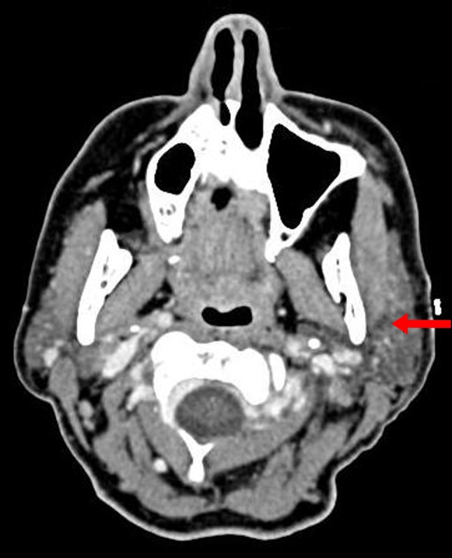
Axial CT image demonstrating a 5 mm left parotid gland node (arrow) deep to the skin marker with surrounding fat stranding
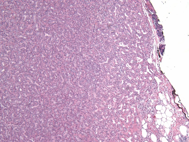
Low magnification view showing a proliferation of small ducts and lacking a well-defined fibrous capsule (H&E, ×100)
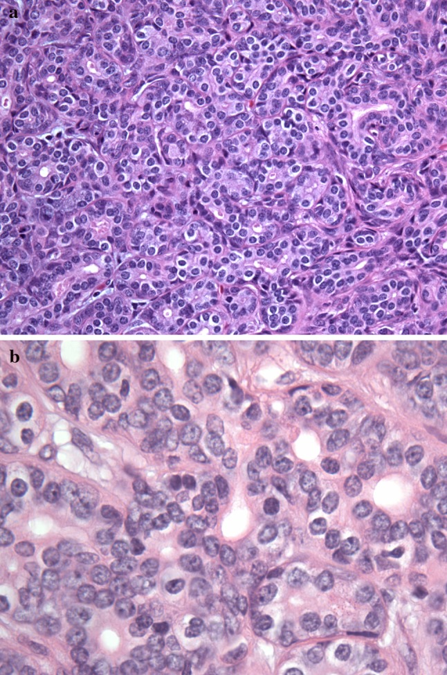
The tubules are composed of a single layer of small, cytologically bland luminal ductal cells with inconspicuous abluminal myoepithelial cells (H&E, a ×400, b ×1,000)
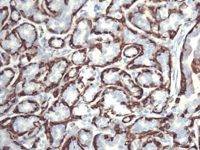
Immunohistochemical staining for calponin highlights an abluminal myoepithelial layer surrounding each small duct (×400)
Similar articles
-
Hybrid Intercalated Duct Lesion of the Parotid: Diagnostic Challenges of a Recently Described Entity with Fine Needle Aspiration Findings.Head Neck Pathol. 2016 Jun;10(2):269-74. doi: 10.1007/s12105-015-0663-8. Epub 2015 Oct 17. Head Neck Pathol. 2016. PMID: 26477034 Free PMC article.
-
Intercalated duct lesions of salivary gland: a morphologic spectrum from hyperplasia to adenoma.Am J Surg Pathol. 2009 Sep;33(9):1322-9. doi: 10.1097/PAS.0b013e3181a55c15. Am J Surg Pathol. 2009. PMID: 19542868
-
A Subset of Salivary Intercalated Duct Lesions Harbors Recurrent CTNNB1 and HRAS Mutations: A Molecular Link to Basal Cell Adenoma and Epithelial-Myoepithelial Carcinoma?Head Neck Pathol. 2023 Jun;17(2):393-400. doi: 10.1007/s12105-022-01513-x. Epub 2022 Dec 8. Head Neck Pathol. 2023. PMID: 36480093 Free PMC article.
-
Hybrid carcinoma of the parotid gland: report of a case (epithelial-myoepithelial carcinoma and salivary duct carcinoma) and review of the literature.Acta Otolaryngol. 2010;130(1):185-9. doi: 10.3109/00016480902930458. Acta Otolaryngol. 2010. PMID: 19449226 Review.
-
Fine-needle aspiration biopsy of alveolar rhabdomyosarcoma of Stensen's duct: a case report and review of the literature.Diagn Cytopathol. 2014 Dec;42(12):1069-74. doi: 10.1002/dc.23084. Epub 2014 Mar 6. Diagn Cytopathol. 2014. PMID: 24599626 Review.
Cited by
-
Parenchymal changes of salivary glands adjacent to a variety of salivary gland disorders.Int J Clin Exp Pathol. 2019 Apr 1;12(4):1124-1133. eCollection 2019. Int J Clin Exp Pathol. 2019. PMID: 31933928 Free PMC article.
-
Top Ten Differentials to Mull Over for Head and Neck Myoepithelial Neoplasms.Head Neck Pathol. 2023 Mar;17(1):1-15. doi: 10.1007/s12105-022-01502-0. Epub 2023 Mar 16. Head Neck Pathol. 2023. PMID: 36928733 Free PMC article. Review.
-
Advances in PSMA theranostics.Transl Oncol. 2022 Aug;22:101450. doi: 10.1016/j.tranon.2022.101450. Epub 2022 May 18. Transl Oncol. 2022. PMID: 35597190 Free PMC article.
-
Hybrid intercalated duct lesion of the parotid: A case report.World J Clin Cases. 2022 Nov 26;10(33):12358-12364. doi: 10.12998/wjcc.v10.i33.12358. World J Clin Cases. 2022. PMID: 36483828 Free PMC article.
-
Salivary gland hybrid tumour revisited: could they represent high-grade transformation in a low-grade neoplasm?Virchows Arch. 2016 Dec;469(6):643-650. doi: 10.1007/s00428-016-2018-6. Epub 2016 Sep 7. Virchows Arch. 2016. PMID: 27605055
References
-
- Elhosseiny A. Salivary glands. In: Koss L, Melamed M, editors. Koss’ diagnostic cytology and its histopathological bases. 5th ed. Lippincott, Philadelphia: Williams & Wilkins; 2006.
Publication types
MeSH terms
LinkOut - more resources
Full Text Sources
