. 2012 Feb 13;209(2):259-73.
doi: 10.1084/jem.20111694. Epub 2012 Jan 23.

Genetic resistance to JAK2 enzymatic inhibitors is overcome by HSP90 inhibition

Affiliations

Genetic resistance to JAK2 enzymatic inhibitors is overcome by HSP90 inhibition

Oliver Weigert et al. J Exp Med. .

Abstract

Enzymatic inhibitors of Janus kinase 2 (JAK2) are in clinical development for the treatment of myeloproliferative neoplasms (MPNs), B cell acute lymphoblastic leukemia (B-ALL) with rearrangements of the cytokine receptor subunit cytokine receptor-like factor 2 (CRLF2), and other tumors with constitutive JAK2 signaling. In this study, we identify G935R, Y931C, and E864K mutations within the JAK2 kinase domain that confer resistance across a panel of JAK inhibitors, whether present in cis with JAK2 V617F (observed in MPNs) or JAK2 R683G (observed in B-ALL). G935R, Y931C, and E864K do not reduce the sensitivity of JAK2-dependent cells to inhibitors of heat shock protein 90 (HSP90), which promote the degradation of both wild-type and mutant JAK2. HSP90 inhibitors were 100-1,000-fold more potent against CRLF2-rearranged B-ALL cells, which correlated with JAK2 degradation and more extensive blockade of JAK2/STAT5, MAP kinase, and AKT signaling. In addition, the HSP90 inhibitor AUY922 prolonged survival of mice xenografted with primary human CRLF2-rearranged B-ALL further than an enzymatic JAK2 inhibitor. Thus, HSP90 is a promising therapeutic target in JAK2-driven cancers, including those with genetic resistance to JAK enzymatic inhibitors.


Figures

Figure 1.
Figure 1.
JAK2 signaling as a therapeutic target. (A) Chemical structure of BVB808. (B) Kinase assays were performed with recombinant kinase (JH1) domains of the respective JAKs to determine the relative JAK-family selectivity of BVB808. (C) BVB808 activity against JAK2-dependent and JAK2-independent cell lines. GI50 values represent means of at least two independent experiments (n = 2–4). (D) JAK2 V617F mutant MB-02 cells were treated with increasing concentrations of BVB808 for 30 min. Inhibition of constitutive pSTAT5 was analyzed by Western blotting using a Tyr694 phospho-specific antibody. Total STAT5 is included as a loading control. (E) JAK2 V617F mutant SET-2 and JAK3 A572V mutant CMK cells were treated with increasing concentrations of BVB808 for 1 h, and then extracted for immunoblotting. (F and G) MB-02 and SET-2 cells were treated with 1 µM BVB808 for up to 24 h. Cell extracts were prepared at different time points as indicated and probed for pSTAT5. Activation of cell death was assessed by detection of cleaved PARP (arrowhead). β-Tubulin was used as a loading control. (H) Efficacy of BVB808 was evaluated in a mouse bone marrow transplant model of Jak2 V617F–driven MPN after 3 wk of dosing. Bar, 100 µm. (I) In a separate experiment, CT imaging of mice before treatment and after 3 and 50 d of vehicle or BVB808. After imaging on day 50, the mice were sacrificed and the spleens were dissected and weighed. Spleen weight is shown on the right. Bar, 1 cm. (J) Immunohistochemistry for pStat5 in spleen and bone marrow sections from samples collected 2 h after the last dose of vehicle or BVB808. Bar, 50 µm.
Figure 2.
Figure 2.
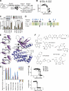
JAK2 alleles that confer resistance to enzymatic inhibitors. (A) In vitro mutagenesis screen of JAK2 R683G in Ba/F3-CRLF2 cells to identify mutations that confer resistance to BVB808. (B) Sensitivity to BVB808 was reduced in Ba/F3-CRFL2/JAK2 R683G cells harboring Y931C (GI50, 303 nM), G935R (462 nM), or E864K (427 nM) compared with no resistance mutation (96.4 nM; P < 0.05 for all 3 mutants). Proliferation was measured based on relative fluorescence units (RFU). Error bars represent SD. (C) Ba/F3 cells expressing ATIC-ALK (ALK+), EpoR/Jak2 V617F alone (none), or EpoR/Jak2 V617F with one of the three kinase domain mutations were transfected with either of two siRNA (#4 and #2) targeting mouse Jak2. The cells were grown in the absence of IL-3, and proliferation was measured after 24 and 48 h and normalized to cells from the same background transfected with nontargeting (nt) control siRNA. Representative immunoblot of VF/Y931C cells 24 h after transfection is shown. (D) Alignment of homologous regions in JAK2 and ABL1. Blue arrows indicate mutated codons in BCR/ABL1 reported to confer imatinib resistance in patients (Soverini et al., 2011). Black arrows indicate codons identified by in vitro mutagenesis of BCR/ABL1 (Azam et al., 2003) and JAK2 E864K, Y931C, and G935R mutations (below). (E) Structure of the kinase domain of JAK2 indicating E864, Y931, and G935 (right), with modeling of wild type, Y931C, and G935R (left, top to bottom). (F) Chemical structures of JAK inhibitors and the HSP90 inhibitor AUY922. (G) E864K, Y931C, and G935R were introduced into Jak2 V617F and expressed in Ba/F3-EpoR cells. Proliferation was assayed 48 h after exposure to indicated drug or vehicle in triplicate on two occasions based on RFU. *, P < 0.05 compared with no resistance mutation for that drug (GI50 in nanomolar in parentheses). (H) Dose response curves based on RFU after 48 h of treatment with indicated drugs. Each data point was obtained in quadruplicate, and the experiment was independently repeated three times. Error bars indicate SD.
Figure 3.
Figure 3.
Functional characterization of resistance mutations in JAK2 V617F. (A) IC50 values of tofacitinib or JAKinh-1 determined using purified wild-type, G935Q, G935H, or G935R JAK2 kinase domains. (B) Ba/F3-EpoR/HA-Jak2 V617F or Ba/F3-EpoR/HA-Jak2 V617F/G935R cells were treated with the indicated drugs for 16 h, and lysates were immunoblotted with antibodies against hemagglutinin (HA) or the indicated proteins. (C) Ba/F3-EpoR cells expressing Jak2 V617F-Thy1.1 and Ba/F3-EpoR cells expressing Jak2 V617F/Y931C-GFP (top)/G935R-GFP (middle)/or E864K-GFP (bottom) were mixed 1:1. The relative fraction of each cell population was quantified by flow cytometry in the presence or absence of drug, as indicated. (D) Mice injected with 1:1 mixes of luciferized Ba/F3-EpoR-JAK2 V617F-Thy1.1 and luciferized Ba/F3-EpoR-Jak2 V617/Y931C-GFP cells were treated with AUY922 (n = 8) or vehicle (n = 9) and luciferase was measured at indicated time points. Error bars indicate standard error of the mean. *, P < 0.05 compared with vehicle. (E) Representative luciferase imaging of AUY922- and vehicle-treated mice on day 8 of treatment. (F) Survival of mice treated with vehicle or AUY922. (G) Peripheral blood was analyzed from mice treated with vehicle (V) or AUY922 (A) by flow cytometry for percentage of mononuclear cells expressing GFP (Jak2 V617F/Y931C) or Thy1.1 (Jak2 V617F). 4 mice were analyzed at each time point except for vehicle day 11 (n = 2). Error bars indicate SEM.
Figure 4.
Figure 4.
JAK2 and HSP90 inhibition in CRLF2-rearranged B-ALL cell lines. (A) MHH-CALL4 and MUTZ-5 cells were exposed to JAK2 enzymatic inhibitors and the HSP90 inhibitor AUY922 at indicated concentrations for 96 h, and proliferation was quantified based on RFU. Error bars represent SD. Shown results are representative of two independent experiments performed in quadruplicate. (B) GI50 concentrations for JAK2 and HSP90 inhibitors after 2 d (Ba/F3 cell lines) or 3 d (MHH-CALL4 and MUTZ-5 cell lines) of exposure. K562 cells harbor BCR/ABL1 and not CRLF2 rearrangements. (C) Immunoblotting in MHH-CALL4 and MUTZ-5 cells exposed to 1 µM JAKinh-1, 50 nM AUY922, combination, or vehicle for 16 h. (D) MHH-CALL4 cells were transfected with siRNA against the indicated JAK family member or with scrambled control, and lysates were collected for immunoblotting after 72 h. (E) Immunoblotting of Ba/F3-CRLF2/JAK2 R683G cells for the indicated total or phosphorylated proteins after treatment with the indicated doses of HSP990 or PU-H71.
Figure 5.
Figure 5.
Transcriptional profiling of JAK2 and HSP90 inhibition in CRLF2-rearranged B-ALL cell lines. MHH-CALL4 and MUTZ-5 cells were treated in triplicate for 14 h with either 1 µM JAKinh-1, 50 nM AUY922, the combination of both (combo), or vehicle. (A) Unsupervised hierarchical clustering of transcriptional profiles. (B) Hierarchical clustering and heat map of the most differentially expressed genes within the top/bottom 20 (FDR < 0.25 and FC > 2.5) between a treatment group and vehicle for MUTZ-5 and MHH-CALL4 cells. More abundant genes are visualized on the colorimetric scale in red, less abundant genes in blue. (C) GSEA was performed for each treatment condition with STAT5A and HSF1 gene signatures. (D) A JAK inhibitor signature was determined from the top/bottom 250 most differentially expressed genes after treatment with JAKinh-1. This signature was used to perform GSEA for comparison AUY922 versus vehicle treatment.
Figure 6.
Figure 6.
JAK2 and HSP90 inhibition in CRLF2-rearranged human xenografts. NSG mice were injected with primary 412 or 537 cells i.v. (n = 40 per line). When leukemia engraftment was >30% in the bone marrow, mice were divided into 4 treatment groups: 50 mg/kg AUY922 i.v. thrice weekly, 50 mg/kg BVB808 by oral gavage twice daily, combination, or vehicle. (A) Immunohistochemistry using H&E or antibodies against human CD45 (hCD45), pSTAT5, or HSP70 in spleens obtained from NSG mice engrafted with 412 leukemias. Mice were treated for 5 d, and spleens were harvested 2–4 h after the last dose. (B) Immunoblotting of lysates from spleens obtained from NSG mice engrafted with 412 or 537 leukemias and then treated for 5 d with the indicated agent(s). (C) Unsupervised hierarchical clustering of transcriptional profiles of 412 or 537 leukemias engrafted in NSG mice after treatment with BVB808, AUY922, BVB808+AUY922 (combination), or vehicle. (D) Survival among NSG mice engrafted with primary human CRLF2-rearranged B-ALL samples and treated with BVB808, AUY922, combination, or vehicle. *, P < 0.05 compared with vehicle; **, P < 0.05 compared with BVB808.

Similar articles

Cited by

References

    1. Abramson J.S., Chen W., Juszczynski P., Takahashi H., Neuberg D., Kutok J.L., Takeyama K., Shipp M.A. 2009. The heat shock protein 90 inhibitor IPI-504 induces apoptosis of AKT-dependent diffuse large B-cell lymphomas. Br. J. Haematol. 144:358–366 10.1111/j.1365-2141.2008.07484.x - DOI - PMC - PubMed
    1. Azam M., Latek R.R., Daley G.Q. 2003. Mechanisms of autoinhibition and STI-571/imatinib resistance revealed by mutagenesis of BCR-ABL. Cell. 112:831–843 10.1016/S0092-8674(03)00190-9 - DOI - PubMed
    1. Baffert F., Régnier C.H., De Pover A., Pissot-Soldermann C., Tavares G.A., Blasco F., Brueggen J., Chène P., Drueckes P., Erdmann D., et al. 2010. Potent and selective inhibition of polycythemia by the quinoxaline JAK2 inhibitor NVP-BSK805. Mol. Cancer Ther. 9:1945–1955 10.1158/1535-7163.MCT-10-0053 - DOI - PubMed
    1. Bareng J., Jilani I., Gorre M., Kantarjian H., Giles F., Hannah A., Albitar M. 2007. A potential role for HSP90 inhibitors in the treatment of JAK2 mutant-positive diseases as demonstrated using quantitative flow cytometry. Leuk. Lymphoma. 48:2189–2195 10.1080/10428190701607576 - DOI - PubMed
    1. Cerchietti L.C., Lopes E.C., Yang S.N., Hatzi K., Bunting K.L., Tsikitas L.A., Mallik A., Robles A.I., Walling J., Varticovski L., et al. 2009. A purine scaffold Hsp90 inhibitor destabilizes BCL-6 and has specific antitumor activity in BCL-6-dependent B cell lymphomas. Nat. Med. 15:1369–1376 10.1038/nm.2059 - DOI - PMC - PubMed

Publication types

MeSH terms