. 2012 Feb;135(Pt 2):447-60.
doi: 10.1093/brain/awr339. Epub 2012 Jan 23.

Transplanted neural stem/precursor cells instruct phagocytes and reduce secondary tissue damage in the injured spinal cord

Affiliations

Transplanted neural stem/precursor cells instruct phagocytes and reduce secondary tissue damage in the injured spinal cord

Melania Cusimano et al. Brain. 2012 Feb.

Abstract

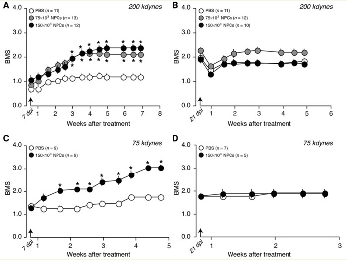
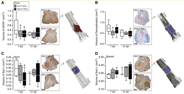
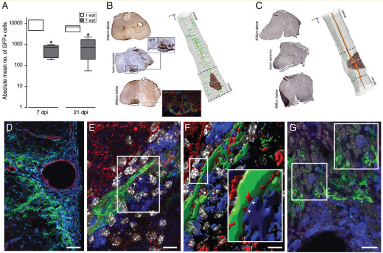
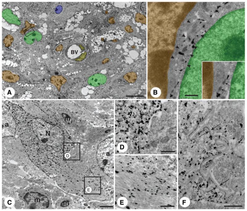
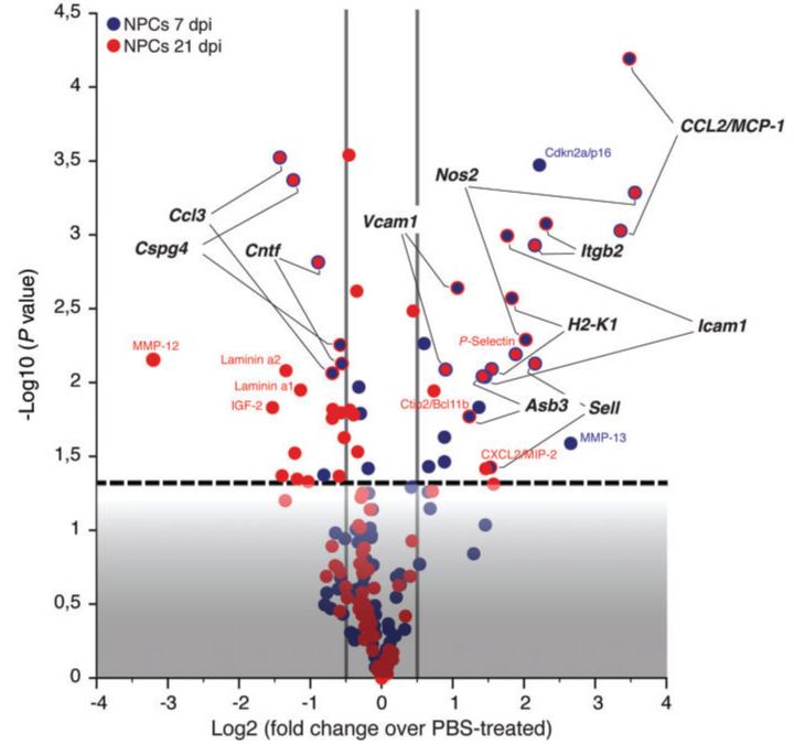
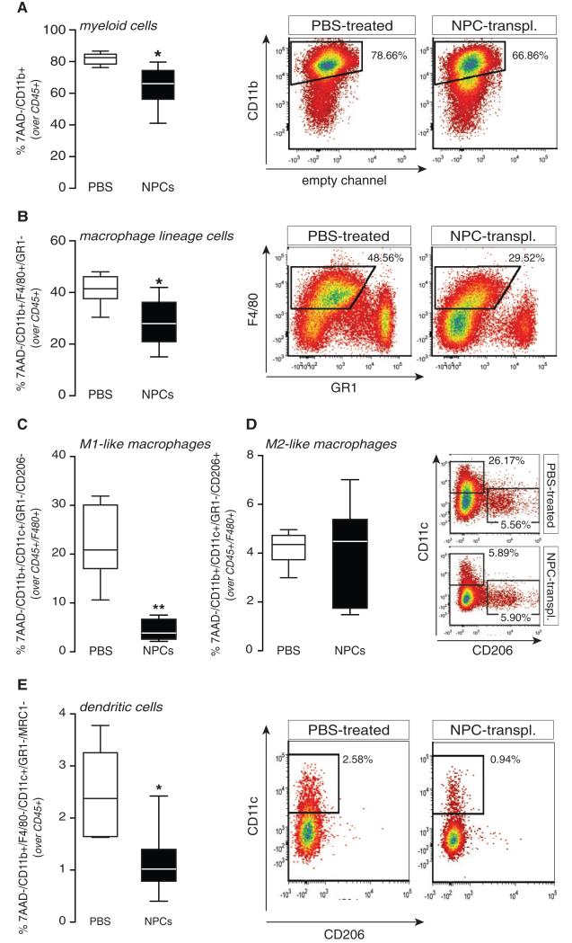
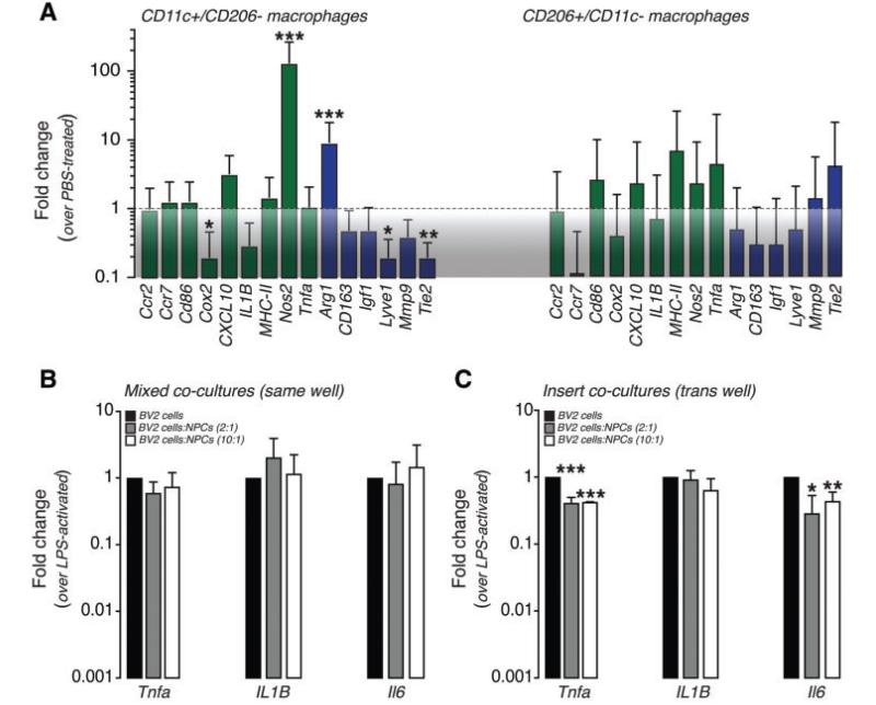
Transplanted neural stem/precursor cells possess peculiar therapeutic plasticity and can simultaneously instruct several therapeutic mechanisms in addition to cell replacement. Here, we interrogated the therapeutic plasticity of neural stem/precursor cells after their focal implantation in the severely contused spinal cord. We injected syngeneic neural stem/precursor cells at the proximal and distal ends of the contused mouse spinal cord and analysed locomotor functions and relevant secondary pathological events in the mice, cell fate of transplanted neural stem/precursor cells, and gene expression and inflammatory cell infiltration at the injured site. We used two different doses of neural stem/precursor cells and two treatment schedules, either subacute (7 days) or early chronic (21 days) neural stem/precursor cell transplantation after the induction of experimental thoracic severe spinal cord injury. Only the subacute transplant of neural stem/precursor cells enhanced the recovery of locomotor functions of mice with spinal cord injury. Transplanted neural stem/precursor cells survived undifferentiated at the level of the peri-lesion environment and established contacts with endogenous phagocytes via cellular-junctional coupling. This was associated with significant modulation of the expression levels of important inflammatory cell transcripts in vivo. Transplanted neural stem/precursor cells skewed the inflammatory cell infiltrate at the injured site by reducing the proportion of 'classically-activated' (M1-like) macrophages, while promoting the healing of the injured cord. We here identify a precise window of opportunity for the treatment of complex spinal cord injuries with therapeutically plastic somatic stem cells, and suggest that neural stem/precursor cells have the ability to re-programme the local inflammatory cell microenvironment from a 'hostile' to an 'instructive' role, thus facilitating the healing or regeneration past the lesion.


Figures

Figure 1
Figure 1
Recovery of locomotor functions after subacute, but not chronic, NPC transplantation in mice with contusion spinal cord injury. Amelioration of locomotor functions by subacutely (A and C), but not chronically (B and D) transplanted NPCs in mice suffering from spinal cord injuries. The arrow indicates the day of treatment. Data are mean Basso Mouse Scale (BMS ± SEM). *P ≤ 0.001, versus PBS-treated controls. dpi = days post-injury.
Figure 2
Figure 2
Promotion of healing in the severely injured spinal cord. (A–D) Stereological quantification of volumes of GFAP+ tissue injury (A; red solid), Luxol fast blue (LFB) demyelination (B; grey solid) and Iba1 + , at the level of the injured (grey solid in C) or spared (grey solid in D) cord tissue. In the 3D renderings, the solid orange in A–D is the central canal, while the solid transparent blue in B–D is the injury volume. Shown in A–D are also representative axial images of the stainings for stereological quantifications. Images have been taken at either 600 μm above the lesion (A, B and D) or at the lesion epicentre (C), as indicated by the dashed lines. Volumes in A–D have been calculated at 56 days after the injury. Data are minimum to maximum volumes from n ≥ 3 mice per group. *P ≤ 0.05, versus PBS-treated controls. dpi = days post-injury; GFAP = glial fibrillary acidic protein.
Figure 3
Figure 3
In vivo survival and integration transplanted NPCs. (A) Quantification of transplanted fGFP+ NPCs in vivo at either 1 or 7 weeks post transplantation (wpt) of 150 × 103 NPCs. Data are absolute minimum to maximum numbers of fGFP+ cells/mouse from n ≥ 3 mice/group. *P ≤ 0.05, as compared with 1 week after transplantation. (B and C) Representative axial images of the GFP staining (brown) for stereological quantifications in A from two representative mice with spinal cord injury sacrificed at 1 (B) or 7 (C) weeks after NPC transplantation. Haematoxylin staining in B and D is shown in blue. The fluorescent image in B shows detail of GFP+ cells at the level of the perivascular area. In the 3D renderings, the red solid is GFAP, the green dots are GFP and the solid orange is the central canal. Dashed lines refer to representative axial images. (D) Confocal microscopy image of an ‘atypical perivascular niche’, where GFP+ NPCs are found in very close vicinity to Iba1+ cells (light blue). CD31+ endothelial cells are in red. (E) Confocal microscopy image of GFP (green) NPCs contacting F4/80+ macrophages via connexin43+ cellular junctions (red; arrowheads). (F) Volocity®-based 3D reconstruction (from a total of n = 20 Z-stacks of optical slices in 0.3 μm intervals) of the confocal Z-stack in E. The magnified inset shows structural junctional connexin43 pattern (red; arrowheads) between the process of one NPC (green) and one juxtaposed F4/80+ macrophage (blue). (G) Confocal microscopy image of GFP (green) NPCs and B220+ putative B lymphocytes (blue) not establishing connexin43+ (red) mediated junctional coupling in a perivascular spinal cord area. DAPI is blue in D and grey in E–G. Scale bars: D = 60 μm; E and F = 15 μm; G = 30 μm.
Figure 4
Figure 4
Morphology and ultrastructure of atypical perivascular NPC niches. (A) Electron micrograph of NPCs labelled with pre-embedding immunogold for GFP. NPCs are highly branched and accumulate at the level of perivascular niches. The main cellular components of these perivascular niches are infiltrating monocyte/macrophages that are identified by their scarce cytoplasm and irregular nucleus with clumped chromatin. The frame indicates one NPC whose processes are found in very close contact to a monocyte/macrophage. (B) High magnification of the frame in A showing the immunogold-labelled process of an NPC (arrowheads) running between a monocyte/macrophage and a second immunogold-labelled NPC. Cellular junctions between both NPC cytoplasms (inset, arrows) and between the NPC and the monocyte/macrophage can be observed in the inset. Pseudo colours in A and B: NPCs = green; monocytes/macrophages = orange; endothelial cells = yellow; endogenous astrocytes = blue. (C) Immunogold-labelled NPC (N) surrounded by two endogenous (immunogold-negative) astrocytes (a), and next to three infiltrating monocytes/macrophages (m). The two frames indicate sites of cell-to-cell contacts. (D) Detail of the contact between the NPC and one endogenous astrocyte (arrowheads). (E) Detail of cytoplasm of the NPC typically rich in intermediate filaments. (F) Detail of an immunogold-positive NPC showing an extremely interdigitated surface surrounded by basal lamina (between arrows). Images in A–F were collected at 50 days after transplantation. Scale bars: A = 5 μm; B = 200 nm; C = 2 μm; D and E = 500 nm; F = 1 μm. BV = blood vessel.
Figure 5
Figure 5
Modulation of gene expression. Volcano plot [x-axis = log2 (fold change); y-axis = Log10 (P-value)] showing statistically significant differentially expressed genes between mice with spinal cord injury transplanted at either 7 days post-injury or 21 days post-injury with 150 × 103 NPCs, as compared with mice with spinal cord injury injected with PBS at the same time points. Vertical grey lines (x = −0.5 and x = 0.5) correspond to fold changes of 0.7 and 1.4, respectively. The horizontal dashed line (y = 1.3) corresponds to a P-value = 0.05. Data have been calculated from n = 5 individual mice per treatment group.
Figure 6
Figure 6
Instruction of professional phagocytes towards a tissue-healing mode. Flow cytometry analysis of myeloid cell subsets in the injured spinal cord at 14 days post-injury (7 days after subacute treatment). CD45+ hematopoietic cells isolated from injured spinal cord were stained with 7-AAD to exclude non-viable cells from further analysis. (A) Myeloid cells; (B) macrophage lineage cells; (C) ‘classically-activated’ (M1-like) inflammatory macrophages; (D) ‘alternatively-activated’ (M2-like) tissue-remodelling/pro-angiogenic macrophages; and (E) dendritic cells. All gates were set based on specific fluorescence minus one (FMO) control samples. For each myeloid cell subset, quantitative data are shown on the left, while representative density plots are shown on the right. White whiskers are mice with spinal cord injury injected with PBS, while black whiskers are spinal cord injury mice injected with 150 × 103 NPCs. Data are minimum to maximum per cent of marker-positive cells from n = 24 mice/treatment group and a total of n = 2 independent experiments. *P ≤ 0.005 and **P ≤ 0.0001, versus PBS-treated controls.
Figure 7
Figure 7
NPCs affect the gene signature of inflammatory professional phagocytes both in vivo and in vitro. (A) Gene signature of spinal cord injury-infiltrating macrophages in NPC-transplanted mice at 14 days post-injury (7 days after subacute treatment). Green bars are markers of M1-like macrophages, whereas blue bars are markers of M2-like macrophages. Data are mean fold changes (over PBS-treated) (± SEM) from n = 24–30 mice/treatment group and n ≥ 2 independent experiments. (B and C) Expression of inflammatory messenger RNAs in BV-2 cells co-cultured with NPCs. Data are mean fold changes (over lipopolysaccharide-activated) (± SEM). *P ≤ 0.05; **P ≤ 0.005 and ***P ≤ 0.0001. LPS = lipopolysaccharide.

Similar articles

Cited by

References

    1. Aharonowiz M, Einstein O, Fainstein N, Lassmann H, Reubinoff B, Ben-Hur T. Neuroprotective effect of transplanted human embryonic stem cell-derived neural precursors in an animal model of multiple sclerosis. PLoS One. 2008;3:e3145. - PMC - PubMed
    1. Alexander JK, Popovich PG. Neuroinflammation in spinal cord injury: therapeutic targets for neuroprotection and regeneration. Prog Brain Res. 2009;175:125–37. - PubMed
    1. Bacigaluppi M, Pluchino S, Peruzzotti-Jametti L, Kilic E, Kilic U, Salani G, et al. Delayed post-ischaemic neuroprotection following systemic neural stem cell transplantation involves multiple mechanisms. Brain. 2009;132:2239–51. - PubMed
    1. Barnabe-Heider F, Frisen J. Stem cells for spinal cord repair. Cell Stem Cell. 2008;3:16–24. - PubMed
    1. Beck KD, Nguyen HX, Galvan MD, Salazar DL, Woodruff TM, Anderson AJ. Quantitative analysis of cellular inflammation after traumatic spinal cord injury: evidence for a multiphasic inflammatory response in the acute to chronic environment. Brain. 2010;133:433–47. - PMC - PubMed

Publication types