

An efficient interlaced multi-shell sampling scheme for reconstruction of diffusion propagators
- PMID: 22271832
- PMCID: PMC3343185
- DOI: 10.1109/TMI.2012.2184551
An efficient interlaced multi-shell sampling scheme for reconstruction of diffusion propagators
Abstract
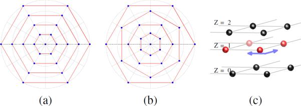
In this paper, we propose an interlaced multi-shell sampling scheme for the reconstruction of the diffusion propagator from diffusion weighted magnetic resonance imaging (DW-MRI). In standard multi-shell sampling schemes, sample points are uniformly distributed on several spherical shells in q-space. The distribution of sample points is the same for all shells, and is determined by the vertices of a selected polyhedron. We propose a more efficient interlaced scheme where sample points are different on alternating shells and are determined by the vertices of a pair of dual polyhedra. Since it samples more directions than the standard scheme, this method offers increased angular discrimination. Another contribution of this work is the application of optimal sampling lattices to q-space data acquisition and the proposal of a model-free reconstruction algorithm, which uses the lattice dependent sinc interpolation function. It is shown that under this reconstruction framework, the body centered cubic (BCC) lattice provides increased accuracy. The sampling scheme and the reconstruction algorithms were evaluated on simulated data as well as rat brain data collected on a 600 MHz (14.1T) Bruker imaging spectrometer.
Figures


Similar articles
-
Single- and Multiple-Shell Uniform Sampling Schemes for Diffusion MRI Using Spherical Codes.IEEE Trans Med Imaging. 2018 Jan;37(1):185-199. doi: 10.1109/TMI.2017.2756072. Epub 2017 Sep 25. IEEE Trans Med Imaging. 2018. PMID: 28952937 Free PMC article.
-
Generalized diffusion spectrum magnetic resonance imaging (GDSI) for model-free reconstruction of the ensemble average propagator.Neuroimage. 2019 Apr 1;189:497-515. doi: 10.1016/j.neuroimage.2019.01.038. Epub 2019 Jan 23. Neuroimage. 2019. PMID: 30684636 Free PMC article.
-
Accelerated multi-shell diffusion MRI with Gaussian process estimated reconstruction of multi-band imaging.Magn Reson Med. 2025 Aug;94(2):694-712. doi: 10.1002/mrm.30518. Epub 2025 Apr 6. Magn Reson Med. 2025. PMID: 40189808 Free PMC article.
-
Impact of radial and angular sampling on multiple shells acquisition in diffusion MRI.Med Image Comput Comput Assist Interv. 2011;14(Pt 2):116-23. doi: 10.1007/978-3-642-23629-7_15. Med Image Comput Comput Assist Interv. 2011. PMID: 21995020
-
Multiple-fiber reconstruction algorithms for diffusion MRI.Ann N Y Acad Sci. 2005 Dec;1064:113-33. doi: 10.1196/annals.1340.018. Ann N Y Acad Sci. 2005. PMID: 16394152 Review.
Cited by
-
Estimating diffusion propagator and its moments using directional radial basis functions.IEEE Trans Med Imaging. 2015 Oct;34(10):2058-78. doi: 10.1109/TMI.2015.2418674. Epub 2015 Mar 31. IEEE Trans Med Imaging. 2015. PMID: 25838518 Free PMC article.
-
Multi-fiber tractography visualizations for diffusion MRI data.PLoS One. 2013 Nov 25;8(11):e81453. doi: 10.1371/journal.pone.0081453. eCollection 2013. PLoS One. 2013. PMID: 24282597 Free PMC article.
-
Tensor metrics and charged containers for 3D Q-space sample distribution.Med Image Comput Comput Assist Interv. 2013;16(Pt 1):679-86. doi: 10.1007/978-3-642-40811-3_85. Med Image Comput Comput Assist Interv. 2013. PMID: 24505726 Free PMC article.
-
Design of multishell sampling schemes with uniform coverage in diffusion MRI.Magn Reson Med. 2013 Jun;69(6):1534-40. doi: 10.1002/mrm.24736. Epub 2013 Apr 26. Magn Reson Med. 2013. PMID: 23625329 Free PMC article.
-
From expected propagator distribution to optimal q-space sample metric.Med Image Comput Comput Assist Interv. 2014;17(Pt 3):217-24. doi: 10.1007/978-3-319-10443-0_28. Med Image Comput Comput Assist Interv. 2014. PMID: 25320802 Free PMC article.
References
-
- Moseley M, Cohen Y, Mintorovitch J, Chileuitt L, Shimizu H, Kucharczyk J, Wendland M, Weinstein P. Early detection of regional cerebral ischemia in cats: comparison of diffusion-and T2-weighted MRI and spectroscopy. Magnetic Resonance in Medicine. 1990;14(2):330–346. - PubMed
-
- Tuch D, Reese T, Wiegell M, Van J W. Diffusion MRI of complex neural architecture. Neuron. 2003;40(5):885–895. - PubMed
-
- Cohen Y, Assaf Y. High b-value q-space analyzed diffusion-weighted MRS and MRI in neuronal tissues-a technical review. NMR in Biomedicine. 2002;15(7–8):516–542. - PubMed
-
- Callaghan P. Principles of Nuclear Magnetic Resonance Microscopy. Clarendon Press; Oxford: 1991.
