. 2012 Mar 15;18(6):1611-8.
doi: 10.1158/1078-0432.CCR-11-1942. Epub 2012 Jan 23.

Constitutive AP-1 activity and EBV infection induce PD-L1 in Hodgkin lymphomas and posttransplant lymphoproliferative disorders: implications for targeted therapy

Affiliations

Constitutive AP-1 activity and EBV infection induce PD-L1 in Hodgkin lymphomas and posttransplant lymphoproliferative disorders: implications for targeted therapy

Michael R Green et al. Clin Cancer Res. .

Erratum in

  • Clin Cancer Res. 2012 Apr 1;18(7):2117

Abstract

Purpose: Programmed cell death ligand 1 (PD-L1) is a molecule expressed on antigen-presenting cells that engages the PD-1 receptor on T cells and inhibits T-cell receptor signaling. The PD-1 axis can be exploited by tumor cells to dampen host antitumor immune responses and foster tumor cell survival. PD-1 blockade has shown promise in multiple malignancies but should be directed toward patients in whom it will be most effective. In recent studies, we found that the chromosome 9p24.1 amplification increased the gene dosage of PD-L1 and its induction by JAK2 in a subset of patients with classical Hodgkin lymphoma (cHL). However, cHLs with normal 9p24.1 copy numbers also expressed detectable PD-L1, prompting analyses of additional PD-L1 regulatory mechanisms.

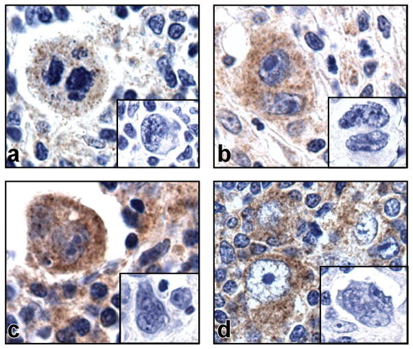
Experimental design: Herein, we utilized immunohistochemical, genomic, and functional analyses to define alternative mechanisms of PD-L1 activation in cHL and additional EBV(+) lymphoproliferative disorders.

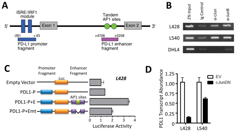
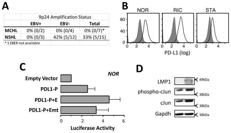
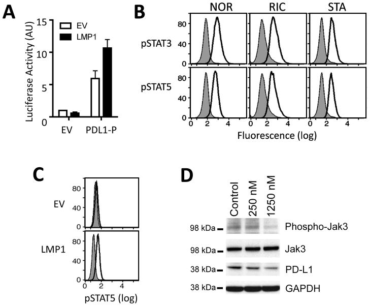
Results: We identified an AP-1-responsive enhancer in the PD-L1 gene. In cHL Reed-Sternberg cells, which exhibit constitutive AP-1 activation, the PD-L1 enhancer binds AP-1 components and increases PD-L1 promoter activity. In addition, we defined Epstein-Barr virus (EBV) infection as an alternative mechanism for PD-L1 induction in cHLs with diploid 9p24.1. PD-L1 was also expressed by EBV-transformed lymphoblastoid cell lines as a result of latent membrane protein 1-mediated, JAK/STAT-dependent promoter and AP-1-associated enhancer activity. In addition, more than 70% of EBV(+) posttransplant lymphoproliferative disorders expressed detectable PD-L1.

Conclusions: AP-1 signaling and EBV infection represent alternative mechanisms of PD-L1 induction and extend the spectrum of tumors in which to consider PD-1 blockade.


Figures

Figure 1
Figure 1. PD-L1 expression in primary cHL tumors
Immunohistochemical staining for PD-L1 on four representative primary cHLs with diploid 9p24.1. PD-L1 expression (brown) is detected in large multinucleated RS cells which are unstained with the isotype control (inset).
Figure 2
Figure 2. Constitutive AP-1 activity supports PD-L1 expression in cHL
A) Diagramatic representation of the PD-L1 regulatory elements, including the previously described ISRE/IRF1 module of the JAK/STAT-responsive promoter and the predicted enhancer element containing dual AP-1 binding sites. Fragments cloned into luciferase constructs are annotated below, with positions relative to the PD-L1 transcription start site. B) Binding of cJUN and JUN-B to the candidate PD-L1 enhancer region following ChiP-coupled PCR. L428 and L540, cHL cell lines; DHL4, DLBCL cell line. Data are representative of two independent experiments. C) PD-L1 promoter and enhancer-driven luciferase activity in the L428 cHL cell line. Constructs include the empty pGL3 vector, the pGL3 vector with the promoter cloned upstream of the luciferase gene alone (PDL1-P), the promoter with the wild-type enhancer cloned downstream of the luciferase gene (PDL1-P+E), and the promoter with an enhancer lacking AP-1 binding sites (PDL1-P+Emt). Data are the average of three independent experiments. D) PD-L1 transcript abundance in cHL cell lines following overexpression of a cJUN dominant-negative (cJUNDN) construct. The effect of cJUNDN expression on PD-L1 abundance was greater in L428, which has lower level 9p24.1 amplification than L540. Data are the average of three independent experiments.
Figure 3
Figure 3. EBV increases PD-L1 expression
A) The frequency of EBV infection and 9p24.1 amplification in a cohort of primary cHL tumors of nodular sclerosis (NSHL) and mixed cellularity (MCHL) subtypes. For each category of primary cHL (NSHL or MCHL, EBV+, EBV or total), the numbers and percentage of 9p24+/all tumors (%) are indicated. EBV infection is mutually exclusive of 9p24.1 amplification. B) Flow cytometric analysis of cell-surface PD-L1 expression in EBV-transformed lymphoblastiod cell lines. Data are representative of three independent experiments. C) PD-L1 promoter- and enhancer-driven luciferase activity in the EBV-transformed NOR LCL. D) Expression of cJUN and phospho-cJUN following LMP1 transfection of 3T3 cells. GAPDH, loading control. Data are representative of three independent experiments.
Figure 4
Figure 4. LMP1 increases PD-L1 promoter activity
A) LMP1-enhanced PD-L1 promoter-driven luciferase activity in 293T cells. 293T cells were cotransfected with empty pGL3 vector (EV) or PDL1-P and the LMP-FLAG expression vector (LMP1) and evaluated for luciferase activity. Data are the average of three independent experiments. B) Intracellular phosphoflow cytometric analysis of phosphorylated STAT3 and STAT5 in EBV-transformed LCLs. Data are representative of three independent experiments. C) Intracellular phosphoflow cytometric analysis of phosphorylated STAT5 in LMP1-transfected 293T cells. EV, empty vector. D) Western analyses of phosphorylated JAK3 and PD-L1 in the NOR-LCL treated with the JAK3 inhibitor VI (250 nM or 1250 nM) or control compound. Total JAK3 and GAPDH, loading controls. Data are representative of 3 independent experiments.
Figure 5
Figure 5. PD-L1 expression in primary PTLD tumors
Immunohistochemical staining for PD-L1 on three representative primary PTLDs. In two of the PTLDs (a and b), PD-L1 expression (brown) is detected in tumor cells with centrocytic/centroblastic morphology which are unstained with isotype control (inset). One tumor shows no staining for PD-L1 (c).

Similar articles

Cited by

References

    1. Kuppers R. The biology of Hodgkin’s lymphoma. Nat Rev Cancer. 2009;9:15–27. - PubMed
    1. Young L, Rickinson A. Epstein-Barr virus: 40 years on. Nat Rev Cancer. 2004;4(10):757–68. - PubMed
    1. Portis T, Dyck P, Longnecker R. Epstein-Barr Virus (EBV) LMP2A induces alterations in gene transcription similar to those observed in Reed-Sternberg cells of Hodgkin lymphoma. Blood. 2002;102(12):4166–78. - PubMed
    1. Vockerodt M, Morgan S, Kuo M, Wei W, Chukwuma M, Arrand J, et al. The Epstein-Barr virus oncoprotein, latent membrane protein-1 reprograms germinal centre B cells towards a Hodgkin’s Reed-Sternberg-like phenotype. J Pathol. 2008;216(1):83–92. - PubMed
    1. Ouyang J, Juszczynski P, Rodig SJ, Green MR, O’Donnell E, Currie T, et al. Viral induction and targeted inhibition of galectin-1 in EBV+ post-transplant lymphoproliferative disorders. Blood. 117:4315–22. accompanying editorial, Blood 2011; 4317:4165–6, cited in SciBx 4314(4319); doi:101038/scibx2011251 2012011. - PubMed

Publication types

MeSH terms

Substances