. 2012 Jul;97(7):1092-100.
doi: 10.3324/haematol.2011.053421. Epub 2012 Jan 22.

Overexpression of FoxM1 offers a promising therapeutic target in diffuse large B-cell lymphoma

Affiliations

Overexpression of FoxM1 offers a promising therapeutic target in diffuse large B-cell lymphoma

Shahab Uddin et al. Haematologica. 2012 Jul.

Abstract

Background: FoxM1 has been shown to play a critical role in the pathogenesis of various epithelial malignancies. However, its role in lymphoid malignancies has not been fully clarified. We, therefore, investigated the role of FoxM1 expression in a large cohort of diffuse large B-cell lymphoma samples and panel of cell lines.

Design and methods: FoxM1 expression was investigated in a large series of diffuse large B-cell lymphoma tissues in a tissue microarray format by immunohistochemistry. Apoptosis was measured by flow cytometry and protein expression was detected by immunoblotting using diffuse large B-cell lymphoma cell lines following treatment with either pharmacological inhibitor of FoxM1 or small interference RNA knockdown strategy. Invasion/migration and soft agar colony assays were also performed following treatment with FoxM1 inhibitor.

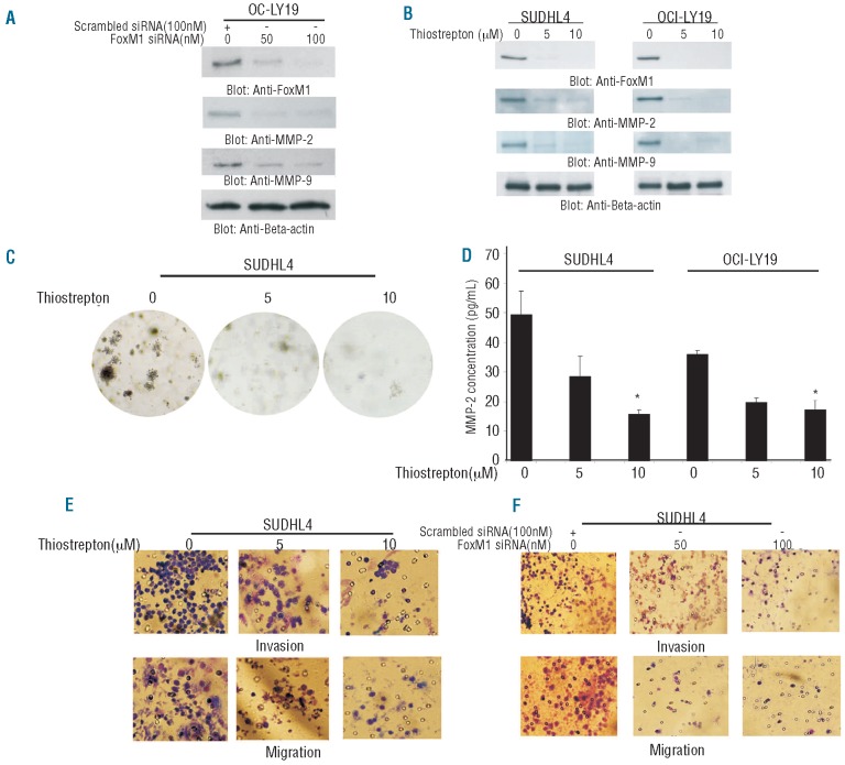
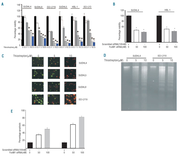
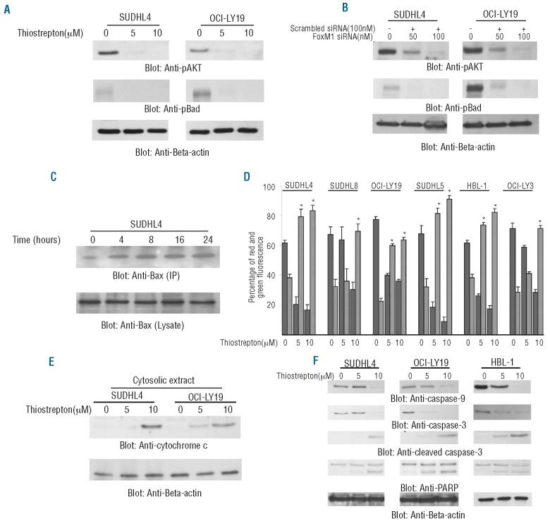
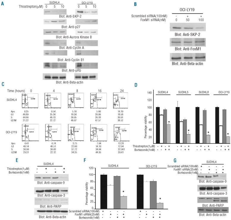
Results: FoxM1 expression was detected in 84.6% of diffuse large B-cell lymphoma tumors and found to be significantly associated with proliferative tumor marker Ki67 (P<0.0001), matrix metalloproteinases-2 (P=0.0008), matrix metalloproteinases-9 (P=0.0002), S-phase kinase associated protein-2 (P<0.0001) and inversely associated with p27 expression (P=0.0215). Expression of small interference RNA targeted against FoxM1 or treatment of diffuse large B-cell lymphoma cells with thiostrepton caused its downregulation accompanied by decreased expression of matrix metalloproteinases-2 and matrix metalloproteinases-9. Inhibition of FoxM1 in diffuse large B-cell lymphoma cells also decreased invasive and migratory capability, and induced caspase dependent apoptosis via activation of the mitochondrial apoptotic pathway. Finally, combined thiostrepton and bortezomib at sub-toxic doses led to efficient apoptosis in diffuse large B-cell lymphoma cells.

Conclusions: Altogether, these results suggest that FoxM1 is over-expressed in the majority of diffuse large B-cell lymphoma samples. These data also indicate that targeting FoxM1 signaling can serve as a potential therapeutic modality in the management of diffuse large B-cell lymphoma.


Figures

Figure 1.
Figure 1.
Thiostrepton treatment causes downregulation of FoxM1 and its downstream targets in DLBCL cells. (A) OCI-LY19 cells were transfected with either 100 nM scrambled siRNA or 50 and 100 nM specific siRNA targeted against FoxM1 for 48 h. After incubation, cells were lysed and immunoblotted with antibodies against FoxM1, MMP-2 and MMP-9. Beta-actin was used as a loading control. (B) SUDHL4 and OCI-LY19 cells were treated with 5 and 10 μM thiostrepton for 48 h. After cell lysis, proteins were separated on SDS-Page and immunoblotted with antibodies against FoxM1, MMP-2, MMP-9 and beta-actin. (C) Clonogenic assays were performed as described in the Design and Methods section. SUDHL4 cells were treated with 5 and 10 μM thiostrepton for 48 h. Subsequently, cells were plated on Soft agar plates for four weeks. After four weeks, plates were stained and manually counted. (D) SUDHL4 and OCI-LY19 cells were treated with 5 and 10 μM thiostrepton for 48 h. Activity of MMP-2 was determined by enzyme-linked immunoabsorbent assay (ELISA). (E) SUDHL4 cells were treated with 5 and 10 μM thiostrepton for 24 h. Following treatment, Invasion-Migration assay were performed as described in the material and methods section. (F) SUDHL4 cells were transfected with either scrambled siRNA or FoxM1 specific siRNA for 48 h. Following treatment, Invasion-Migration assay were performed as described in the Design and Methods section.
Figure 2.
Figure 2.
Downregulation of FoxM1 expression leads to inhibition of cell viability and induction of apoptosis in DLBCL cell lines. (A) DLBCL cells were incubated with 0.5, 1, 5, 10 and 25 μM thiostrepton for 48 h. Cell viability was assayed using MTT as described in Design and Methods section. The graph displays the mean ± SD (standard deviation) of 3 independent experiments with replicates of 6 wells for all the doses and vehicle control for each experiment * P<0.05, statistically significant (Student’s t-test.) (B) SUDHL4 and HBL-1 cells were transfected with scrambled siRNA or FoxM1 specific siRNA for 48 h. Following treatment, cell viability was measured by MTT. The graph displays the mean ± SD (standard deviation) of 3 independent experiments with replicates of 6 wells for all the doses and vehicle control for each experiment *P<0.05, statistically significant (Student’s t-test.) (C) SUDHL4, SUDHL5, SUDHL8 and OCI-LY19 cells were treated with 5 and 10 μM thiostrepton for 48 h and apoptosis was measured by Live/Dead Assay. (D) Thiostrepton-induced apoptosis detected by DNA laddering. DLBCL cells were treated with 5 and 10 μM thiostrepton (as indicated) for 48 h and DNA was extracted and separated by electrophoresis on 1.5% agarose gel. (E) SUDHL4 and HBL-1 cells were transfected with scrambled siRNA or FoxM1 specific siRNA for 48 h. Following treatment, cells were subsequently stained with flourescein-conjugated annexin-V and propidium iodide (PI) and analyzed by flow cytometry. Bar graph denotes standard deviation of 3 independent transfections.
Figure 3.
Figure 3.
Thiostrepton-induced mitochondrial apoptotic pathway in DLBCL cells. (A) SUDHL4 and OCI-LY19 cells were treated with 5 and 10 μM thiostrepton for 48 h and cells were lysed and equal amounts of proteins were separated by SDS-PAGE, transferred to PVDF membrane, and immunoblotted with antibodies against p-AKT, p-Bad and Beta-actin. (B) SUDHL4 and OCI-LY19 cells were transfected with either 100 nM scrambled siRNA or 50 and 100 nM specific siRNA targeted against FoxM1 for 48 h. After incubation, cells were lysed and immunoblotted with antibodies against p-AKT, p-Bad and Beta-actin. (C) After treating with 10 μM thiostrepton for indicated time periods, SUDHL4 cells were lysed in 1% Chaps lysis buffer and subjected to immunoprecipitation with anti-Bax 6A7 monoclonal antibody and probed with specific polyclonal anti-Bax antibody for detection of conformationally changed Bax protein. In addition, the total cell lysates were applied directly to SDS–PAGE, transferred to immobilon membrane and immunoblotted with specific anti-Bax polyclonal antibody. (D) Loss of mitochondrial membrane potential by thiostrepton treatment of DLBCL cells. DLBCL cells were treated with and without 5 and 10 μM thiostrepton for 48 h. Live cells with intact mitochondrial membrane potential and dead cells with lost mitochondrial membrane potential were measured by JC-1 staining and analyzed by flow cytometry as described in Design and Methods section. The graph displays the mean ± SD (standard deviation) of 3 independent experiments. (E) Thiostrepton-induced release of cytochrome c. SUDHL4 and OCI-LY19 cells were treated with and without 5 and 10 μM thiostrepton for 48 h. Mitochondrial free, cytosolic fractions were isolated as described in Design and Methods section. Cell extracts were separated on SDS-PAGE, transferred to PVDF membrane, and immunoblotted with an antibody against cytochrome c. The blots were stripped and re-probed with an antibody against actin for equal loading. (F) SUDHL4, OCI-LY19 and HBL-1 cells were treated with and without 5 and 10 μM thiostrepton for 48 h. Cells were lysed and equal amounts of proteins were separated by SDS-PAGE, transferred to PVDF membrane, and immunoblotted with antibodies against caspase-9, caspase-3, cleaved caspase-3, PARP and beta-actin.
Figure 4.
Figure 4.
Association of FoxM1 expression with cell cycle regulatory proteins. (A) SUDHL4 and OCI-LY19 cells were treated with and without 5 and 10 μM thiostrepton for 48 h. After cell lysis, equal amounts of proteins were separated by SDS-PAGE, transferred to Immobilon membrane, and immunoblotted with antibodies against SKP-2, p27Kip1, Aurora kinase B, Cyclin A, Cyclin B1, pRb and beta-actin as indicated. A representative of 3 different experiments is shown. (B) SKP-2 siRNA expression does not affect expression of FoxM1. OCI-LY19 cells were transfected with scrambled siRNA (100 nM) and SKP-2 siRNA (50 and 100 nM) with Lipofectamine as described in the Design and Methods section. After 48 h of transfection, cells were lysed and equal amounts of proteins were separated by SDS-PAGE, transferred to Immobilon membrane, and immunoblotted with antibodies against SKP-2, FoxM1 and beta-actin as indicated. (C) Thiostrepton treatment induces a G2/M cell cycle arrest at early time point in DLBCL. SUDHL4 and OCI-LY19 cells were treated with 10 μM thiostrepton for indicated time periods. Following incubation, cells were analyzed for cell cycle fractions by flow cytometry. (D) DLBCL cells were incubated with either 1 μM thiostrepton or 1nM bortezomib alone or in combination for 48 h. Cell viability was assayed using MTT as described in the Design and Methods section. The graph displays the mean ± SD (standard deviation) of 3 independent experiments with replicates of 6 wells for all the doses and vehicle control for each experiment *P<0.05, statistically significant (Student’s t-test.). (E) SUDHL4 cells were treated with either 1 μM thiostrepton or 1 nM bortezomib alone or in combination (as indicated) for 48 h. Cells were lysed and equal amounts of proteins were separated by SDS-PAGE, transferred to PVDF membrane, and immunoblotted with antibodies against caspase-9, caspase-3, PARP and beta-actin. (F) SUDHL4 and OCI-LY19 cells were trans-fected with either 100 nM scrambled siRNA or 25 nM specific siRNA targeted against FoxM1 alone or in combination with 1 nM bortezomib for 48 h. Cell viability was assayed using MTT as described in Design and Methods section. The graph displays the mean ± SD (standard deviation) of 3 independent experiments with replicates of 6 wells for all the doses and vehicle control for each experiment. (G) SUDHL4 cells were transfected with either 100 nM scrambled siRNA or 25 nM specific siRNA targeted against FoxM1 alone or in combination with 1 nM bortezomib for 48 h. Cells were lysed and equal amounts of proteins were separated by SDS-PAGE, transferred to PVDF membrane, and immunoblotted with antibodies against caspase-9, caspase-3, PARP and beta-actin.

Similar articles

Cited by

References

    1. Escalon MP, Lossos IS. Pharmacotherapy of large B-cell lymphoma. Expert Opin Pharmacother. 2008;9(13):2247–58. - PubMed
    1. Fisher RI. Cyclophosphamide, doxorubicin, vincristine, and prednisone versus intensive chemotherapy in non-Hodgkin’s lymphoma. Cancer Chemother Pharmacol. 1997;40(Suppl):S42–6. - PubMed
    1. Elenitoba-Johnson KS, Jenson SD, Abbott RT, Palais RA, Bohling SD, Lin Z, et al. Involvement of multiple signaling pathways in follicular lymphoma transformation: p38-mitogen-activated protein kinase as a target for therapy. Proc Natl Acad Sci USA. 2003;100(12):7259–64. - PMC - PubMed
    1. Markovic O, Marisavljevic D, Cemerikic V, Perunicic M, Savic S, Filipovic B, et al. Clinical and prognostic significance of apoptotic profile in patients with newly diagnosed nodal diffuse large B-cell lymphoma (DLBCL) Eur J Haematol. 2011;86(3):246–55. - PubMed
    1. Staudt LM, Dave S. The biology of human lymphoid malignancies revealed by gene expression profiling. Adv Immunol. 2005;87:163–208. - PMC - PubMed

MeSH terms