Altered immunophenotypic features of peripheral blood platelets in myelodysplastic syndromes
- PMID: 22271903
- PMCID: PMC3366656
- DOI: 10.3324/haematol.2011.057158
Altered immunophenotypic features of peripheral blood platelets in myelodysplastic syndromes
Abstract
Background: Multiparameter flow cytometric analysis of bone marrow and peripheral blood cells has proven to be of help in the diagnostic workup of myelodysplastic syndromes. However, the usefulness of flow cytometry for the detection of megakaryocytic and platelet dysplasia has not yet been investigated. The aim of this pilot study was to evaluate by flow cytometry the diagnostic and prognostic value of platelet dysplasia in myelodysplastic syndromes.
Design and methods: We investigated the pattern of expression of distinct surface glycoproteins on peripheral blood platelets from a series of 44 myelodysplastic syndrome patients, 20 healthy subjects and 19 patients with platelet alterations associated to disease conditions other than myelodysplastic syndromes. Quantitative expression of CD31, CD34, CD36, CD41a, CD41b, CD42a, CD42b and CD61 glycoproteins together with the PAC-1, CD62-P, fibrinogen and CD63 platelet activation-associated markers and platelet light scatter properties were systematically evaluated.
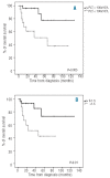
Results: Overall, flow cytometry identified multiple immunophenotypic abnormalities on platelets of myelodysplastic syndrome patients, including altered light scatter characteristics, over-and under expression of specific platelet glycoproteins and asynchronous expression of CD34; decreased expression of CD36 (n = 5), CD42a (n = 1) and CD61 (n = 2), together with reactivity for CD34 (n = 1) were only observed among myelodysplastic syndrome cases, while other alterations were also found in other platelet disorders. Based on the overall platelet alterations detected for each patient, an immunophenotypic score was built which identified a subgroup of myelodysplastic syndrome patients with a high rate of moderate to severe alterations (score>1.5; n = 16) who more frequently showed thrombocytopenia, megakaryocytic dysplasia and high-risk disease, together with a shorter overall survival.
Conclusions: Our results show the presence of altered phenotypes by flow cytometry on platelets from around half of the myelodysplastic syndrome patients studied. If confirmed in larger series of patients, these findings may help refine the diagnostic and prognostic assessment of this group of disorders.
Figures


References
-
- Greenberg PL, Young NS, Gattermann N. Myelodysplastic syndromes. Hematology Am Soc Hematol Educ Program. 2002:136–61. - PubMed
-
- Tefferi A, Vardiman JW. Myelodysplastic syndromes. N Engl J Med. 2009;361(19):1872–85. - PubMed
-
- Brunning RD, Orazi A, Germing U, Le Beau MM, Porwit A, Baumann I, et al. Chapter 5, Myelodysplastic syndromes/neoplasms overview. Lyon: IARC; 2008. WHO classification of tumours of haematopoietic and lymphoid tissues; pp. 88–93.
-
- Valent P, Horny HP, Bennett JM, Fonatsch C, Germing U, Greenberg P, et al. Definitions and standards in the diagnosis and treatment of the myelodysplastic syndromes: Consensus statements and report from a working conference. Leuk Res. 2007;31(6):727–36. - PubMed
-
- Loken MR, van de Loosdrecht A, Ogata K, Orfao A, Wells DA. Flow cytometry in myelodysplastic syndromes: report from a working conference. Leuk Res. 2008;32(1):5–17. - PubMed
Publication types
MeSH terms
Substances
LinkOut - more resources
Full Text Sources
Medical
Miscellaneous

