

Spontaneous Cardiac Hypertrophy in a Crl:CD(SD) Rat
- PMID: 22271980
- PMCID: PMC3246022
- DOI: 10.1293/tox.22.83
Spontaneous Cardiac Hypertrophy in a Crl:CD(SD) Rat
Abstract
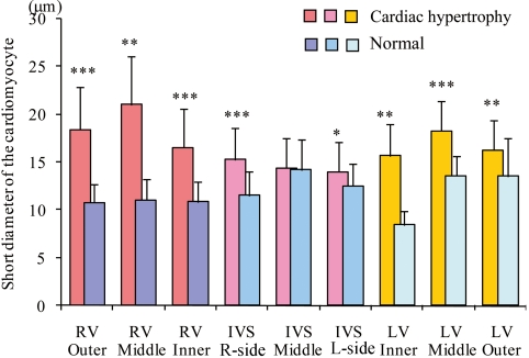
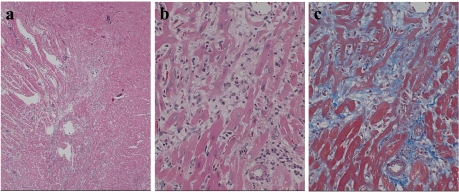
Cardiac hypertrophy was observed in a 9-week-old Crl:CD(SD) rat that died unexpectedly. The animal was allocated to the control group of a toxicity study, and no abnormalities in its general conditions, body weight or food intake were observed. Necropsy revealed an increase in heart weight. Gross examination indicated cardiac enlargement with thickening of the right and left ventricular walls. Histopathological examination revealed hypertrophy of the cardiomyocytes in the right and left ventricular walls and the interventricular septum. Electron microscopic examination indicated bizarre nuclei and accumulation of an increased number of various sizes of mitochondria in the perinuclear region of the hypertrophied myocytes. Hypertrophied myocytes connected by intensely folded intercalated disks were also observed. Based on these findings, the animal was diagnosed with cardiac hypertrophy. This is the first case report of cardiac hypertrophy in this strain.
Keywords: SD rat; heart; hypertrophy.
Figures


Similar articles
-
Myocyte cellular hypertrophy is responsible for ventricular remodelling in the hypertrophied heart of middle aged individuals in the absence of cardiac failure.Cardiovasc Res. 1994 Aug;28(8):1199-208. doi: 10.1093/cvr/28.8.1199. Cardiovasc Res. 1994. PMID: 7954623
-
[Interconnection of parameters of the mitochondrial and myofibrillar apparatus of cardiomyocytes and ploidy and hypertrophy in certain mammalian species, differing in body mass].Tsitologiia. 1997;39(10):946-64. Tsitologiia. 1997. PMID: 9505342 Russian.
-
Aging, cardiac hypertrophy and ischemic cardiomyopathy do not affect the proportion of mononucleated and multinucleated myocytes in the human heart.J Mol Cell Cardiol. 1996 Jul;28(7):1463-77. doi: 10.1006/jmcc.1996.0137. J Mol Cell Cardiol. 1996. PMID: 8841934
-
Spontaneous hypertrophic cardiomyopathy in a cynomolgus macaque (Macaca fascicularis).J Toxicol Pathol. 2018 Jan;31(1):49-54. doi: 10.1293/tox.2017-0027. Epub 2017 Oct 22. J Toxicol Pathol. 2018. PMID: 29479140 Free PMC article.
-
[Role of the cardiac renin-angiotensin system in hypertensive heart disease].Herz. 1995 Oct;20(5):322-9. Herz. 1995. PMID: 7498879 Review. German.
Cited by
-
Establishment of a model of spontaneously-running-Tokushima-shikoku rats with left atrial thrombosis.J Toxicol Pathol. 2014 Apr;27(1):51-6. doi: 10.1293/tox.2012-0032. Epub 2014 Apr 30. J Toxicol Pathol. 2014. PMID: 24791067 Free PMC article.
References
-
- Doggrell SA, Brown L. Rat models of hypertension, cardiac hypertrophy and failure. Cardiovasc Res. 39: 89–105 1998 - PubMed
-
- Hasenfuss G. Animal models of human cardiovascular disease, heart failure and hypertrophy. Cardiovasc Res. 39: 60–76 1998 - PubMed
-
- Mercadier JJ, Lompre AM, Wisnewsky C, Samuel JL, Bercovici J, Swynghedauw B, Schwartz K. Myosin isoenzyme changes in several models of rat cardiac hypertrophy. Circ Res. 49: 525–532 1981 - PubMed
-
- Okamoto K, Aoki K, Nosaka S, Fukushima M. Cardiovascular diseases in the spontaneously hypertensive rat. Jpn Circ J. 28: 943–952 1964 - PubMed
-
- Aiello EA, Villa-Abrille MC, Escudero EM, Portiansky EL, Perez NG, Hurtado MCC, Cingolani HE. Myocardial hypertrophy of normotensive Wistar-Kyoto rats. Am J Physiol Heart Circ Physiol. 286: H1229–H1235 2003 - PubMed
LinkOut - more resources
Full Text Sources