

Spontaneous iron accumulation in hepatocytes of a 7-week-old female rat
- PMID: 22271995
- PMCID: PMC3252042
- DOI: 10.1293/tox.22.199
Spontaneous iron accumulation in hepatocytes of a 7-week-old female rat
Abstract
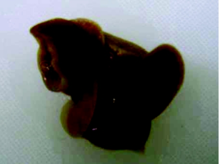
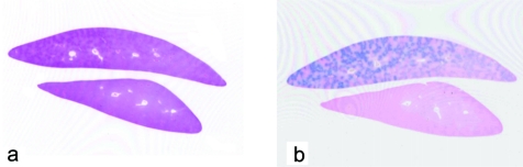
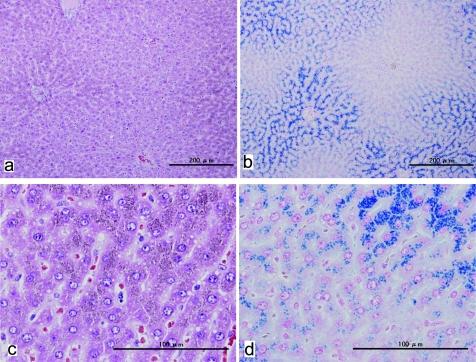
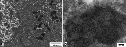
Spontaneous iron accumulation in hepatocytes was observed in a 7-week-old female Han Wistar GALAS rat. Very fine yellowish brown pigments, which showed a positive reaction with Berlin Blue stain, were apparent in the cytoplasm close to the bile canaliculi, with a diminishing periportal-to-centrilobular gradient. There were also differences in distribution between and within lobes. Transmission electron microscopy revealed cytosolic ferritin and pericanalicular siderosomes in hepatocytes. No degeneration or necrotic changes were observed, and non-hepatocyte cells did not demonstrate any obvious accumulation of iron. There were no abnormalities in the animal other than this finding in the liver.
Keywords: hepatocyte; iron accumulation; iron overload; rats; spontaneous.
Figures
Similar articles
-
Ultrastructural sequences during liver iron overload in genetic hemochromatosis.J Hepatol. 1997 Oct;27(4):628-38. doi: 10.1016/s0168-8278(97)80079-7. J Hepatol. 1997. PMID: 9365038 Clinical Trial.
-
Spontaneous iron overload in Sprague-Dawley rats.Toxicol Pathol. 1997 May-Jun;25(3):308-16. doi: 10.1177/019262339702500308. Toxicol Pathol. 1997. PMID: 9210262
-
Pathologic changes in the cytokeratin pericanalicular sheath in experimental cholestasis and alcoholic fatty liver.Lab Invest. 1988 Jul;59(1):60-74. Lab Invest. 1988. PMID: 2455832
-
Iron overload of the liver in the baboon. An ultrastructural study.J Hepatol. 1985;1(3):261-75. doi: 10.1016/s0168-8278(85)80054-4. J Hepatol. 1985. PMID: 4067258
-
Effects of iron and copper overload on the human liver: an ultrastructural study.Curr Med Chem. 2014;21(33):3768-74. doi: 10.2174/0929867321666140601163244. Curr Med Chem. 2014. PMID: 24934354 Review.
Cited by
-
A novel rat model of hereditary hemochromatosis due to a mutation in transferrin receptor 2.Comp Med. 2013 Apr;63(2):143-55. Comp Med. 2013. PMID: 23582421 Free PMC article.
-
Background Data for General Toxicology Parameters in RccHan:WIST Rats at 8, 10, 19 and 32 Weeks of Age.J Toxicol Pathol. 2011 Dec;24(4):195-205. doi: 10.1293/tox.24.195. Epub 2012 Jan 7. J Toxicol Pathol. 2011. PMID: 22319231 Free PMC article.
References
-
- Batts KP. Iron overload syndromes and the liver. Modern Pathology. 20: S31–S39 2007 - PubMed
-
- Iancu TC, Deugnier Y, Halliday JW, Powell LW, Brissot P. Ultrastructural sequences during liver iron overload in genetic hemochromatosis. J Hepatol. 27: 628–638 1997 - PubMed
-
- Masson R, Roome NO. Spontaneous iron overload in Sprague-Dawley rats. Toxicol Pathol. 25: 308–316 1997 - PubMed
-
- Ward RJ, Florence AL, Baldwin D, Abiaka C, Roland F, Ramsey MH, Dickson DP, Peters TJ, Crichton RR. Biochemical and biophysical investigations of the ferrocene-iron-loaded rat. An animal model of primary haemochromatosis. Eur J Biochem. 202: 405–410 1991 - PubMed
-
- Whittaker P, Hines FA, Robl MG, Dunkel VC. Histopathological evaluation of liver, pancreas, spleen, and heart from iron-overloaded Sprague-Dawley rats. Toxicol Pathol. 24: 558–563 1996 - PubMed
LinkOut - more resources
Full Text Sources