

Lesions in the Larynx of Wistar RccHan: WIST Rats
- PMID: 22271998
- PMCID: PMC3234598
- DOI: 10.1293/tox.22.229
Lesions in the Larynx of Wistar RccHan: WIST Rats
Abstract
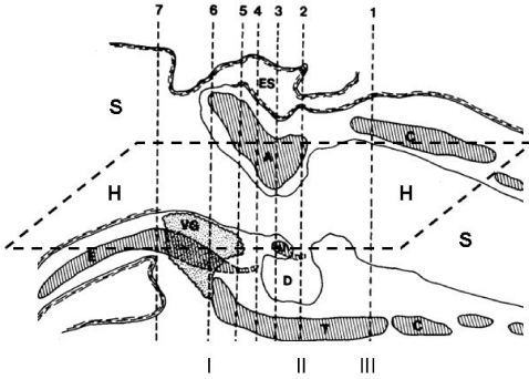
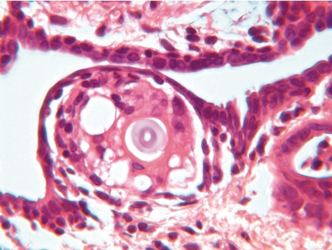
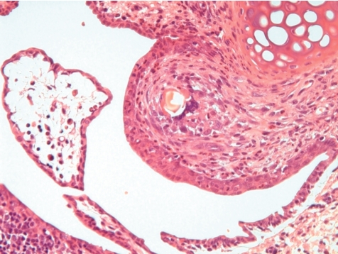
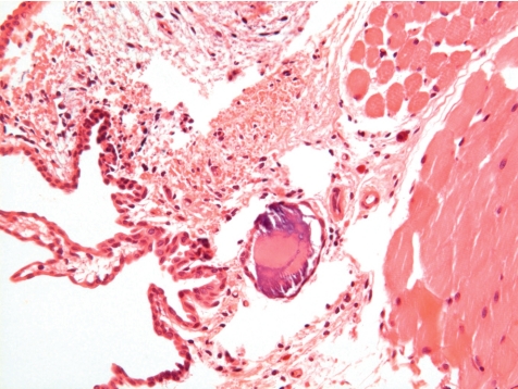
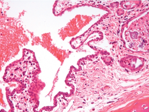
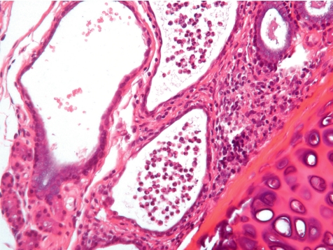
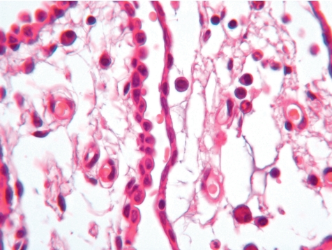
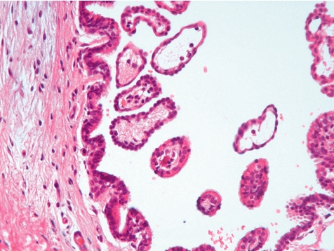
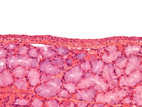
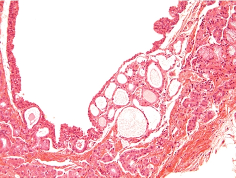
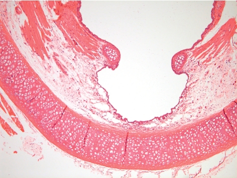
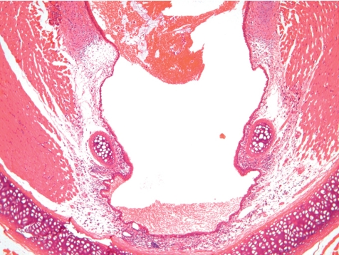
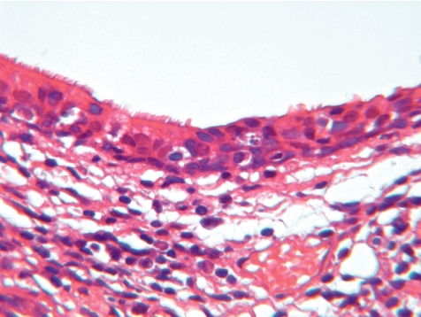
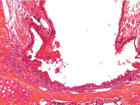
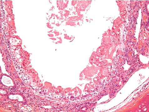
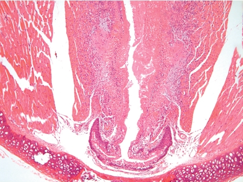
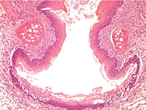
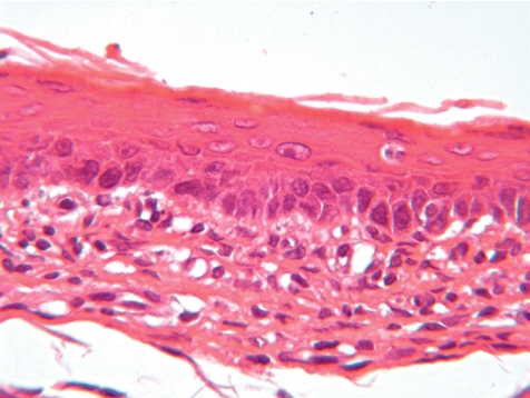
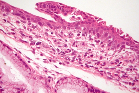
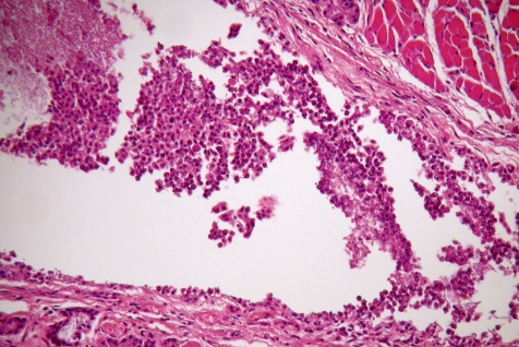
Specific regions in the rat larynx exhibit cellular changes in response to inhaled xenobiotics. These regions include the base of the epiglottis, ventral pouch, and medial surfaces of the vocal processes of the arytenoid cartilages. 1 , 2 In order to collect information on the usefulness of trimming techniques, the influence of different vehicles, the impact of different application routes in toxicity studies, and differences between induced vs. spontaneous lesions, the data obtained from a large number of inhalation and non-inhalation studies performed in Wistar RCCHan(TM): Wist rats at Harlan Laboratories Ltd Switzerland, all evaluated or reviewed by the same pathologist, were compiled for a detailed review. The value of different trimming techniques was deemed to be greatest for transverse and sagittolongitudinal section techniques, as compared to horizontolongitudinally section techniques. The comparison of lesions encountered in control rats of inhalation studies treated with different vehicles did not reveal differences in the type, distribution pattern, incidence and/or severity of spontaneous lesions. The types of lesions were also independent of different application routes in non-inhalation studies compared to inhalation studies. The pattern of spontaneous lesions in the rodent larynx was determined by degenerative and inflammatory lesions starting most often in the submucosal glands by desiccated secretion followed by mineralization and local inflammation or were induced by impacted foreign bodies. Squamous metaplasia was recorded in the respiratory epithelium overlaying the ventral gland as a spontaneous lesion in male Wistar rats from inhalation studies with a maxim of 20.0% in an inhalation oncogenicity study. Induced metaplastic changes recorded in the larynx were reversible. Other induced lesions in inhalation studies consisted of submucosal edema, necrosis, inflammation and/or granuloma. Induced lesions in non-inhalation studies were found to be exclusively related to reflux laryngitis or food impaction. It is concluded, that in rodents induced lesions of the larynx differ in type, distribution pattern, severity and incidence from spontaneous lesions.
Keywords: Wistar RccHanTM: WIST; induced laryngeal lesions; spontaneous laryngeal lesions.
Figures
Similar articles
-
NTP Toxicology and Carcinogenesis Studies of Ozone (CAS No. 10028-15-6) and Ozone/NNK (CAS No. 10028-15-6/ 64091-91-4) in F344/N Rats and B6C3F1 Mice (Inhalation Studies).Natl Toxicol Program Tech Rep Ser. 1994 Oct;440:1-314. Natl Toxicol Program Tech Rep Ser. 1994. PMID: 12595923
-
A histopathological analysis of spontaneous neoplastic and non-neoplastic lesions in aged male RccHan:WIST rats.J Toxicol Pathol. 2020 Jan;33(1):47-55. doi: 10.1293/tox.2019-0064. Epub 2019 Nov 22. J Toxicol Pathol. 2020. PMID: 32051666 Free PMC article.
-
Histologic methods and interspecies variations in the laryngeal histology of F344/N rats and B6C3F1 mice.Toxicol Pathol. 1992;20(1):44-51. doi: 10.1177/019262339202000106. Toxicol Pathol. 1992. PMID: 1411130
-
A Comparison of Rodent and Nonrodent Laryngeal and Tracheal Bifurcation Sensitivities in Inhalation Toxicity Studies and Their Relevance for Human Exposure.Toxicol Pathol. 2017 Jan;45(1):216-222. doi: 10.1177/0192623316678695. Epub 2016 Nov 23. Toxicol Pathol. 2017. PMID: 27879438 Review.
-
Toxicologic significance of histologic change in the larynx of the rat following inhalation exposure: a critical review.Toxicol Appl Pharmacol. 2007 Dec 15;225(3):229-37. doi: 10.1016/j.taap.2007.08.027. Epub 2007 Sep 12. Toxicol Appl Pharmacol. 2007. PMID: 17991503 Review.
Cited by
-
Research-Relevant Conditions and Pathology of Laboratory Mice, Rats, Gerbils, Guinea Pigs, Hamsters, Naked Mole Rats, and Rabbits.ILAR J. 2021 Dec 31;62(1-2):77-132. doi: 10.1093/ilar/ilab022. ILAR J. 2021. PMID: 34979559 Free PMC article. Review.
References
-
- Renne RA, Gideon KM, Miller RA, Mellick PW, Grumbein SL. Histologic methods and interspecies variations in the laryngeal histology of F344/N rats and B6C3F1 mice. Toxicol Pathol. 1992;20:44–51. - PubMed
-
- Renne RA, Sagartz JW, Burger GT. Interspecies variations in the histology of toxicologically important areas in the larynges of CRL:CD rats and Syrian golden hamsters. Toxicol Pathol. 1993;21:542–546. - PubMed
-
- Renne RA, Gideon KM. Types and patterns of response in the larynx following inhalation. Toxicol Pathol. 2006;34:281–285. - PubMed
-
- Renne RA, Gideon KM, Harbo SJ, Staska LM, Grumbein SL. Upper respiratory tract lesions in inhalation toxicology. Toxicol Pathol. 2007;35:163–169. - PubMed
-
- Sagartz JW, Madarasz AJ, Forsell MA, Burger GT, Ayres PH, Coggins CRE. Histological sectioning of the rodent larynx for inhalation toxicity testing. Toxicol Pathol. 1992;20:118–121. - PubMed
LinkOut - more resources
Full Text Sources