. 2010 Sep;23(3):133-9.
doi: 10.1293/tox.23.133. Epub 2010 Oct 5.

Morphological and Biochemical Changes During Aging and Photoaging of the Skin of C57BL/6J Mice

Affiliations

Morphological and Biochemical Changes During Aging and Photoaging of the Skin of C57BL/6J Mice

Ayako Sayama et al. J Toxicol Pathol. 2010 Sep.

Abstract

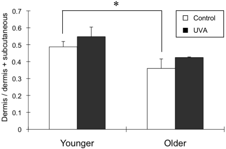
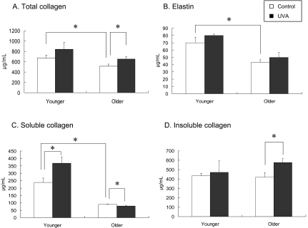
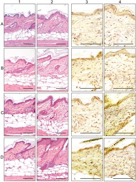
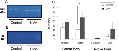
The differences between the dorsal skin of 11- and 16-week-old C57BL/6J mice were examined morphologically and biochemically. The dermis of the 16-week-old mice was thinner than that of the 11-week-old mice due to decreases in the amounts of soluble collagen and elastin. Next, the changes in dorsal skin exposed to UVA irradiation for 8 weeks (576 J/cm(2)) were examined in 3 (younger)- and 8 (older)-week-old C57BL/6J mice. The thickness of the dermis was not significantly different between the UVA-irradiated and control mice in either the younger or older group. The increase in the amount of collagen was related to the increase in the level of soluble collagen in the younger mice. In contrast, it was related to the increase in the level of insoluble collagen in the older mice. In the UVA-irradiated older mice, the activity of the latent form of MMP-13 was significantly higher than that in the control mice. These results suggest that aging and UVA-induced photoaging in the skin are histologically and biochemically different phenomena.

Keywords: UVA; aging; mouse; photoaging; skin.


Figures

Fig. 1
Fig. 1
Relative thickness of the dermis in the 8-week non- and UVA-irradiated mice. The relative thickness of the dermis (the ratio of the dermis to the dermis plus subcutaneous adipose tissue) was calculated at 8 weeks after with and without UVA irradiation in both the younger and older mice (n=3). In the non-irradiated mice, the relative thickness of the dermis in the older mice was lower than that in the younger mice (*P<0.05). UVA-irradiated mice showed an increase in the relative thickness of the dermis in each group, but the difference was not significant.
Fig. 2
Fig. 2
The amounts of dermal collagen and elastin in the 8-week non- and UVA-irradiated mice. The amount of total collagen, which consists of soluble and insoluble collagen, and that of elastin in the skin were measured at 8 weeks after with and without UVA irradiation in both the younger and older mice (n=3). In the non-irradiated mice, the amounts of total collagen, soluble collagen and elastin in the older mice were significantly lower than those in the younger mice (*P<0.05; A, B, and C). The amount of insoluble collagen was similar in the younger and older mice (D). In the UVA-irradiated mice, the amounts of total collagen and elastin tended to be higher than those in the non-irradiated control mice in both groups (A and B). The amount of soluble collagen in the UVA-irradiated mice was significantly higher than that in the non-irradiated control mice in the younger group (*P<0.05; C). The amount of insoluble collagen in the UVA-irradiated mice was also significantly higher than that in the non-irradiated control mice in the older group (*P<0.05; D).
Fig. 3
Fig. 3
Histological changes in the skin of older mice exposed to 2 (A), 4 (B), 6 (C) and 8 (D) weeks of UVA irradiation (columns 2 and 4) and the non-irradiated controls (columns 1 and 3). (columns 1 and 2): H&E staining. Inflammatory cell infiltration into the dermis at 2 weeks after irradiation (A-2), hyperkeratosis in the epidermis at 4 weeks after irradiation (B-2), thickening of the dermis and increased amount of collagen fibers in the subcutaneous layer at 8 weeks after irradiation (D-2). (columns 3 and 4): Weigert’s elastic fiber staining. Increased number of elastic fibers in the dermis at 4 weeks after irradiation (B-4) and tangle formation of elastic fibers at 6 weeks after irradiation (C-4). Bar=50 μm.
Fig. 4
Fig. 4
MMP-13 activity in 8-week UVA-irradiated mice. The results of zymographic analysis of the younger (A) and older (B) UVA-irradiated mice. The bands at 60 and 48 kDa indicate latent and active MMP-13, respectively. The latent form of MMP-13 activity in the UVA-irradiated mice was significantly higher than that of the non-irradiated control mice (*P<0.05) in the older group (C).

References

    1. Kadunce DP, Burr R, Gress R, Kanner R, Lyon JL, Zone JJ. Cigarette smoking: risk factor for premature facial wrinkling. Ann Intern Med. 1991;114:840–844. - PubMed
    1. Placzec M, Kerkmann U, Bell S, Koepke P, Przybilla B. Tobacco smoke is phototoxic. Br J Dermatol. 2004;150:991–993. - PubMed
    1. Fisher GJ, Kang S, Varani J, Bata-Csorgo Z, Wan Y, Datta S, Voorhees J. Mechanisms of photoaging and chronological skin aging. Arch Dermatol. 2002;138:1462–1470. - PubMed
    1. Landau M. Exogenous factors in skin aging. Curr Probl Dermatol. 2007;35:1–13. - PubMed
    1. Kligman LH. The hairless mouse. Model for photoaging. Clin Dermatol. 1996;14:183–195. - PubMed