

Pulmonary fibrosis in response to environmental cues and molecular targets involved in its pathogenesis
- PMID: 22272040
- PMCID: PMC3234628
- DOI: 10.1293/tox.24.9
Pulmonary fibrosis in response to environmental cues and molecular targets involved in its pathogenesis
Abstract
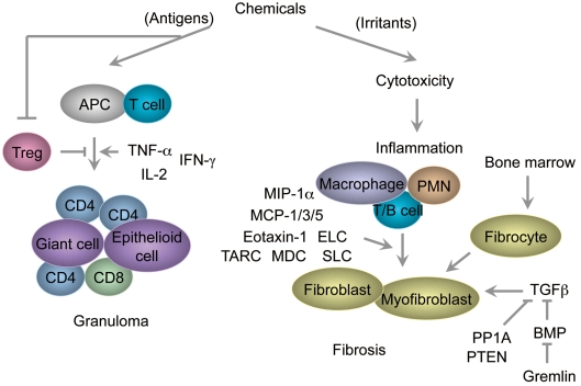
Chronic lung injury resulting from a variety of different causes is frequently associated with the develop ment of pulmonary fibrosis in humans. Although the etiology of pulmonary fibrosis is generally unknown, several sources of evidence support the hypothesis that a number of environmental and occupational agents play an etiologic role in the pathogenesis of this disease. The agents discussed in this review include beryllium, nylon flock, textile printing aerosols, polyvinyl chloride and didecyldimethylammonium chloride. The authors also describe a variety of animal models, including genetically modified mice, in order to investigate the molecular mechanism of pulmonary fibrosis, focusing on chemokine receptors, regulatory T cells and transforming growth factor-β and bone morphogenetic protein signaling. Overall, we propose the concept of toxicological pulmonary fibrosis as a lung disease induced in response to environmental cues.
Keywords: DDAC; TGF-β; beryllium; chemokine receptor; nylon flock; regulatory T cell.
Figures
Similar articles
-
Didecyldimethylammonium chloride induces pulmonary fibrosis in association with TGF-β signaling in mice.Exp Toxicol Pathol. 2013 Nov;65(7-8):1003-9. doi: 10.1016/j.etp.2013.02.003. Epub 2013 Mar 26. Exp Toxicol Pathol. 2013. PMID: 23537712
-
Didecyldimethylammonium chloride induces pulmonary inflammation and fibrosis in mice.Exp Toxicol Pathol. 2010 Nov;62(6):643-51. doi: 10.1016/j.etp.2009.08.007. Epub 2009 Sep 16. Exp Toxicol Pathol. 2010. PMID: 19762220
-
Epithelium-specific deletion of TGF-β receptor type II protects mice from bleomycin-induced pulmonary fibrosis.J Clin Invest. 2011 Jan;121(1):277-87. doi: 10.1172/JCI42090. Epub 2010 Dec 6. J Clin Invest. 2011. PMID: 21135509 Free PMC article.
-
Cytokines and pulmonary fibrosis.Biol Signals. 1996 Jul-Aug;5(4):232-9. doi: 10.1159/000109195. Biol Signals. 1996. PMID: 8891199 Review.
-
Role of Microbial Agents in Pulmonary Fibrosis .Yale J Biol Med. 2017 Jun 23;90(2):219-227. eCollection 2017 Jun. Yale J Biol Med. 2017. PMID: 28656009 Free PMC article. Review.
Cited by
-
TRPM7 restrains plasmin activity and promotes transforming growth factor-β1 signaling in primary human lung fibroblasts.Arch Toxicol. 2022 Oct;96(10):2767-2783. doi: 10.1007/s00204-022-03342-x. Epub 2022 Jul 21. Arch Toxicol. 2022. PMID: 35864199 Free PMC article.
-
Immunization with recombinant Pb27 protein reduces the levels of pulmonary fibrosis caused by the inflammatory response against Paracoccidioides brasiliensis.Mycopathologia. 2015 Feb;179(1-2):31-43. doi: 10.1007/s11046-014-9815-8. Epub 2014 Dec 9. Mycopathologia. 2015. PMID: 25487973
-
Public Health Risks, Dermatological Manifestations, and Environmental Justice Associated With Vinyl Chloride Exposure: Narrative Review.JMIR Dermatol. 2023 Sep 7;6:e48998. doi: 10.2196/48998. JMIR Dermatol. 2023. PMID: 37676716 Free PMC article. Review.
-
Low let-7d exosomes from pulmonary vascular endothelial cells drive lung pericyte fibrosis through the TGFβRI/FoxM1/Smad/β-catenin pathway.J Cell Mol Med. 2020 Dec;24(23):13913-13926. doi: 10.1111/jcmm.15989. Epub 2020 Nov 12. J Cell Mol Med. 2020. PMID: 33179861 Free PMC article.
-
The clinical and immunologic features of pulmonary fibrosis in sarcoidosis.Transl Res. 2012 Nov;160(5):321-31. doi: 10.1016/j.trsl.2012.03.005. Epub 2012 Apr 10. Transl Res. 2012. PMID: 22683422 Free PMC article.
References
-
- Greaves P. Respiratory tract. In: Histopathology of Preclinical Toxicity Studies: Interpretation and Relevance in Drug Safety Evaluation, Third Edition. Elsevier Inc., Oxford, UK. 215–269. 2007
-
- King TE., JrClinical advances in the diagnosis and therapy of the interstitial lung diseases. Am J Respir Crit Care Med. 172: 268–279 2005 - PubMed
-
- American Thoracic Society European Respiratory Society American Thoracic Society/European Respiratory Society International Multidisciplinary Consensus Classification of the Idiopathic Interstitial Pneumonias(This joint statement of the American Thoracic Society (ATS), and the European Respiratory Society (ERS) was adopted by the ATS board of directors, June 2001 and by the ERS Executive Committee, June 2001). Am J Respir Crit Care Med. 165: 277–304 2002 - PubMed
-
- Maher TM, Wells AU, Laurent GJ. Idiopathic pulmonary fibrosis: multiple causes and multiple mechanisms? Eur Respir J. 30: 835–839 2007 - PubMed
-
- Visscher DW, Myers JL. Histologic spectrum of idiopathic interstitial pneumonias. Proc Am Thorac Soc. 3: 322–329 2006 - PubMed
LinkOut - more resources
Full Text Sources