. 2012:2012:683065.
doi: 10.1155/2012/683065. Epub 2012 Jan 5.

In Vitro and In Vivo Antitumor Effect of Anti-CD33 Chimeric Receptor-Expressing EBV-CTL against CD33 Acute Myeloid Leukemia

Affiliations

In Vitro and In Vivo Antitumor Effect of Anti-CD33 Chimeric Receptor-Expressing EBV-CTL against CD33 Acute Myeloid Leukemia

A Dutour et al. Adv Hematol. 2012.

Abstract

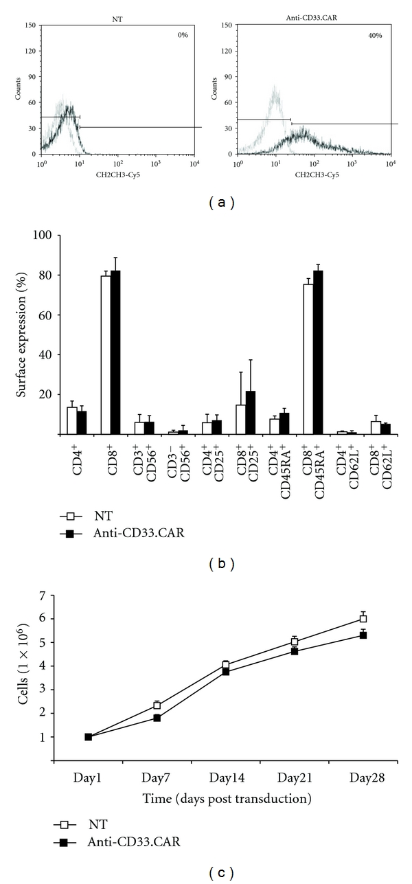
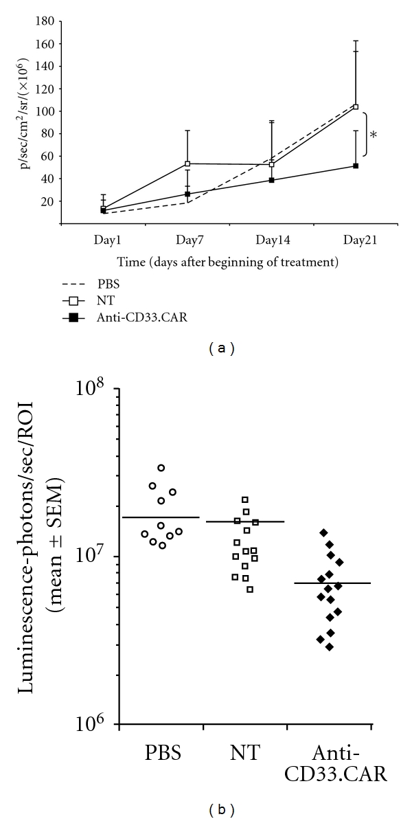
Genetic engineering of T cells with chimeric T-cell receptors (CARs) is an attractive strategy to treat malignancies. It extends the range of antigens for adoptive T-cell immunotherapy, and major mechanisms of tumor escape are bypassed. With this strategy we redirected immune responses towards the CD33 antigen to target acute myeloid leukemia. To improve in vivo T-cell persistence, we modified human Epstein Barr Virus-(EBV-) specific cytotoxic T cells with an anti-CD33.CAR. Genetically modified T cells displayed EBV and HLA-unrestricted CD33 bispecificity in vitro. In addition, though showing a myeloablative activity, they did not irreversibly impair the clonogenic potential of normal CD34(+) hematopoietic progenitors. Moreover, after intravenous administration into CD33(+) human acute myeloid leukemia-bearing NOD-SCID mice, anti-CD33-EBV-specific T cells reached the tumor sites exerting antitumor activity in vivo. In conclusion, targeting CD33 by CAR-modified EBV-specific T cells may provide additional therapeutic benefit to AML patients as compared to conventional chemotherapy or transplantation regimens alone.


Figures

Figure 1
Figure 1
EBV-CTLs could be stably transduced with the anti-CD33.CAR without alteration in their native immunophenotype and expansion rate. (a) The expression of the anti-CD33.CAR on the surface of EBV-CTLs was evaluated by flow cytometry with a Cy5-conjugated-mAb specific for the CH2CH3 domain of the CAR after 7 days of culture. A representative plot of EBV-CTLs transduction after 7 days of culture is shown. (b) The expression of CD4, CD8, CD3 along with CD56, CD4, and CD8 along with CD25, CD4, and CD8 along with CD45RA, CD4, and CD8 along with CD62L on the surface of EBV-CTLs was evaluated after 30 days of culture by flow cytometry. (c) Proliferation of anti-CD33.CAR-transduced EBV-CTLs compared to unmanipulated EBV-CTLs was evaluated by cell count with Trypan blue exclusion after weekly stimulations at 4 : 1 ratio with either irradiated autologous LCLs. Cells were cultured with low-dose rhIL-2 (20 U/mL). Data shown are mean ± SD of 6 separate experiments.
Figure 2
Figure 2
Anti-CD33.CAR-transduced EBV-CTLs efficiently killed autologous LCLs and CD33+ KG-1 cells. Cytotoxicity of unmanipulated EBV-CTLs and anti-CD33.CAR-transduced EBV-CTLs was evaluated by a standard 4-hour 51Chromium-release assay after 7 days of culture at effector:target (E : T) ratios of 50 : 1, 25 : 1, 10 : 1 and 5 : 1 against autologous LCLs (a) and CD33+ KG-1 cell line (b). To assess the specificity of either HLA-I-mediated and CAR-induced killing, target cells were preincubated with anti-human HLA-I (a) antibodies or an anti-human CD33 monoclonal antibody (b) before the addition of the effector cells. Data shown are mean ± SD of 6 separate experiments; *P ≤ 0.05.
Figure 3
Figure 3
Anti-CD33.CAR-transduced EBV-CTLs released substantial levels of IFN-γ and Granzyme B when stimulated with either autologous LCLs and CD33+ KG-1 cells. Release of IFN-γ (a) and Granzyme B (b) from unmanipulated or anti-CD33.CAR-transduced EBV-CTLs was evaluated by ELISpot assays after stimulation with either irradiated autologous LCLs or irradiated KG-1 cells at 1 : 2 ratio. To assess the specificity of either HLA-I-mediated and CAR-induced cytokine release target cells were preincubated with anti-human HLA-I antibodies or an anti-human CD33 monoclonal antibody before the addition of the effector cells. Data shown are mean ± SD of 6 separate experiments; *P ≤ 0.05; **P ≤ 0.005.
Figure 4
Figure 4
Cytotoxicity of anti-CD33.CAR-transduced EBV-CTLs against normal myeloid progenitors. (a) Unmanipulated or anti-CD33.CAR-transduced EBV-CTLs were incubated with bone-marrow-derived CD34+ progenitors at E : T ratio of 1 : 1 for 4 hours. Cells were then seeded in methylcellulose-based medium and after 10 days CFU-GM, CFU-GEMM, and BFU-E were counted. Data shown are mean ± SD of 6 independent experiments. (b) Colonies were collected and characterized by flow cytometry to assess the expression of surface markers specific for each colony type. Data shown are mean ± SD of 6 independent experiments.
Figure 5
Figure 5
In vivo activity of anti-CD33.CAR-transduced EBV-CTLs against AML10. NOD/SCID mice were subcutaneously injected with ffLUC+AML10 cells, and after 5 days mice were weekly intravenously injected with either PBS, unmanipulated or anti-CD33.CAR-transduced EBV-CTLs. (a) Tumor volume was monitored weekly after D-luciferin injection using a Living Image software and was quantified as total photon flux normalized for exposure time and surface area and expressed in units of photons (p) per second per cm2 per steradian (sr). Data shown are mean ± SD of 8 mice/group; *P ≤ 0.05. (b) Intensity signals were log-transformed and summarized using mean ± SD at baseline and multiple subsequent time points for each group of mice. Changes in intensity of signal from baseline at each time point were calculated.
Figure 6
Figure 6
Infiltration of tumors by EBV-CTLs. Forty-eight hours after i.v. administration of EBV-CTLs, tumors were resected, fixed in 10% formalin solution, and embedded in paraffin. CD8+ EBV-CTLs were detected by immunohistochemistry using an anti-CD8 antibody and reported in (a) unmodified EBV-CTLs, (b) CD33-EBV-CTLs. Genetically modified T cells were found at the periphery of the tumor in the vicinity of vessels (indicated as “V”), isolated or forming cluster (arrowheads or arrows, resp.).

Similar articles

Cited by

References

    1. Kowolik CM, Topp MS, Gonzalez S, et al. CD28 costimulation provided through a CD19-specific chimeric antigen receptor enhances in vivo persistence and antitumor efficacy of adoptively transferred T cells. Cancer Research. 2006;66(22):10995–11004. - PubMed
    1. Pieper M, Scheffold C, Duwe S, et al. Immunotherapy of B-cell malignancies with genetically engineered human CD8+ natural killer T cells. Leukemia. 2006;20(4):729–732. - PubMed
    1. Ahmed N, Ratnayake M, Savoldo B, et al. Regression of experimental medulloblastoma following transfer of HER2-specific T cells. Cancer Research. 2007;67(12):5957–5964. - PubMed
    1. Vera J, Savoldo B, Vigouroux S, et al. T lymphocytes redirected against the κ light chain of human immunoglobulin efficiently kill mature B lymphocyte-derived malignant cells. Blood. 2006;108(12):3890–3897. - PMC - PubMed
    1. Faulkner GC, Krajewski AS, Crawford DH. The ins and outs of EBV infection. Trends in Microbiology. 2000;8(4):185–189. - PubMed