

Recent advances in morphological cell image analysis
- PMID: 22272215
- PMCID: PMC3261466
- DOI: 10.1155/2012/101536
Recent advances in morphological cell image analysis
Abstract
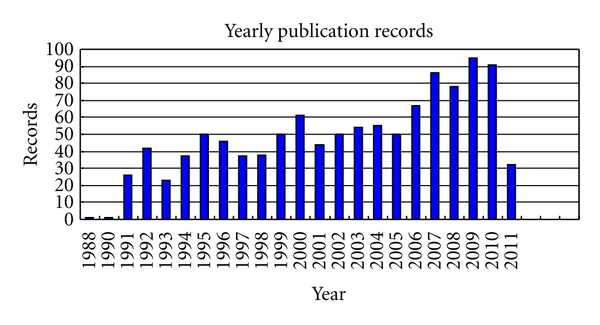
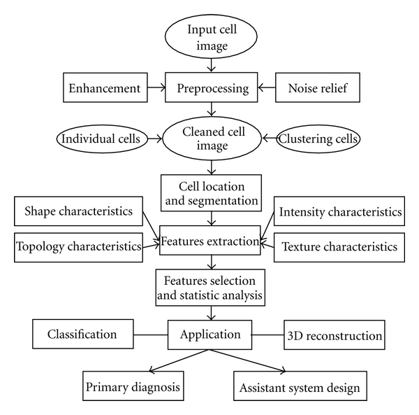
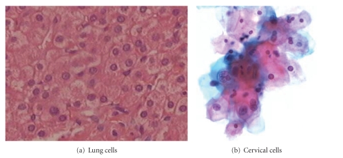
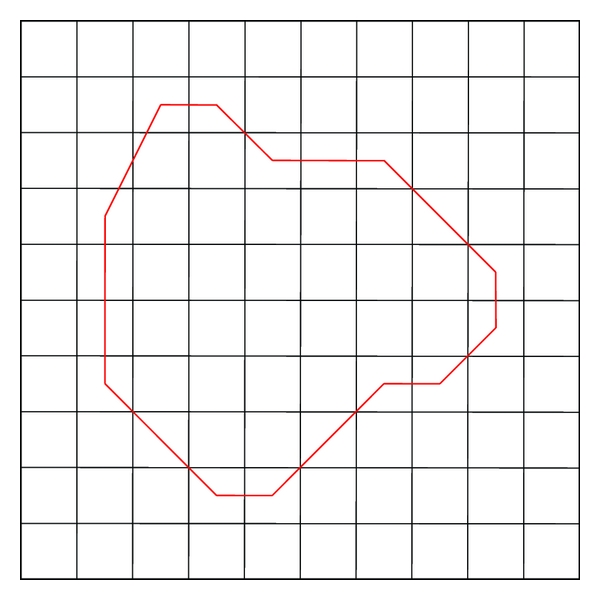
This paper summarizes the recent advances in image processing methods for morphological cell analysis. The topic of morphological analysis has received much attention with the increasing demands in both bioinformatics and biomedical applications. Among many factors that affect the diagnosis of a disease, morphological cell analysis and statistics have made great contributions to results and effects for a doctor. Morphological cell analysis finds the cellar shape, cellar regularity, classification, statistics, diagnosis, and so forth. In the last 20 years, about 1000 publications have reported the use of morphological cell analysis in biomedical research. Relevant solutions encompass a rather wide application area, such as cell clumps segmentation, morphological characteristics extraction, 3D reconstruction, abnormal cells identification, and statistical analysis. These reports are summarized in this paper to enable easy referral to suitable methods for practical solutions. Representative contributions and future research trends are also addressed.
Figures
Similar articles
-
[Review: Segmentation and classification methods of 3D medical images].Zhongguo Yi Liao Qi Xie Za Zhi. 2002 Mar;26(3):197-206. Zhongguo Yi Liao Qi Xie Za Zhi. 2002. PMID: 16104307 Review. Chinese.
-
A review on biomedical image processing and future trends.Comput Methods Programs Biomed. 1990 Mar-Apr;31(3-4):141-83. doi: 10.1016/0169-2607(90)90001-p. Comput Methods Programs Biomed. 1990. PMID: 2194742 Review.
-
Motion analysis of live objects by super-resolution fluorescence microscopy.Comput Math Methods Med. 2012;2012:859398. doi: 10.1155/2012/859398. Epub 2011 Nov 17. Comput Math Methods Med. 2012. PMID: 22162725 Free PMC article. Review.
-
[Study on statistical method of distribution for erythrocyte morphological features by computerized image processing].Sheng Wu Yi Xue Gong Cheng Xue Za Zhi. 2000 Dec;17(4):429-32, 443. Sheng Wu Yi Xue Gong Cheng Xue Za Zhi. 2000. PMID: 11211832 Chinese.
-
Integration of morphological preprocessing and fractal based feature extraction with recursive feature elimination for skin lesion types classification.Comput Methods Programs Biomed. 2019 Sep;178:201-218. doi: 10.1016/j.cmpb.2019.06.018. Epub 2019 Jun 16. Comput Methods Programs Biomed. 2019. PMID: 31416550
Cited by
-
Theorems and application of local activity of CNN with five state variables and one port.Comput Math Methods Med. 2012;2012:674243. doi: 10.1155/2012/674243. Epub 2012 Apr 12. Comput Math Methods Med. 2012. PMID: 22611440 Free PMC article.
-
Estradiol Induces Epithelial to Mesenchymal Transition of Human Glioblastoma Cells.Cells. 2020 Aug 21;9(9):1930. doi: 10.3390/cells9091930. Cells. 2020. PMID: 32825553 Free PMC article.
-
A rate-distortion-based merging algorithm for compressed image segmentation.Comput Math Methods Med. 2012;2012:648320. doi: 10.1155/2012/648320. Epub 2012 Oct 15. Comput Math Methods Med. 2012. PMID: 23118800 Free PMC article.
-
Nonlocal means-based denoising for medical images.Comput Math Methods Med. 2012;2012:438617. doi: 10.1155/2012/438617. Epub 2012 Feb 20. Comput Math Methods Med. 2012. PMID: 22454694 Free PMC article.
-
Profiling DNA damage in 3D Histology Samples.Med Opt Imaging Virtual Microsc Image Anal (2022). 2022 Sep 15:84-93. doi: 10.1007/978-3-031-16961-8_9. Online ahead of print. Med Opt Imaging Virtual Microsc Image Anal (2022). 2022. PMID: 39899002 Free PMC article.
References
-
- Reyes-Aldasoro CC, Williams LJ, Akerman S, Kanthou C, Tozer GM. An automatic algorithm for the segmentation and morphological analysis of microvessels in immunostained histological tumour sections. Journal of Microscopy. 2011;242(3):262–278. - PubMed
-
- Cheng JZ, Chou YH, Huang CS, et al. ACCOMP: augmented cell competition algorithm for breast lesion demarcation in sonography. Medical Physics. 2010;37(12):6240–6252. - PubMed
-
- Schildkraut JS, Prosser N, Savakis A, et al. Level-set segmentation of pulmonary nodules in megavolt electronic portal images using a CT prior. Medical Physics. 2010;37(11):5703–5710. - PubMed
-
- Plissiti ME, Nikou C, Charchanti A. Combining shape, texture and intensity features for cell nuclei extraction in Pap smear images. Pattern Recognition Letters. 2011;32(6):838–853.
Publication types
MeSH terms
LinkOut - more resources
Full Text Sources
