. 2012;7(1):e29021.
doi: 10.1371/journal.pone.0029021. Epub 2012 Jan 17.

Genome-wide progesterone receptor binding: cell type-specific and shared mechanisms in T47D breast cancer cells and primary leiomyoma cells

Affiliations

Genome-wide progesterone receptor binding: cell type-specific and shared mechanisms in T47D breast cancer cells and primary leiomyoma cells

Ping Yin et al. PLoS One. 2012.

Abstract

Background: Progesterone, via its nuclear receptor (PR), exerts an overall tumorigenic effect on both uterine fibroid (leiomyoma) and breast cancer tissues, whereas the antiprogestin RU486 inhibits growth of these tissues through an unknown mechanism. Here, we determined the interaction between common or cell-specific genome-wide binding sites of PR and mRNA expression in RU486-treated uterine leiomyoma and breast cancer cells.

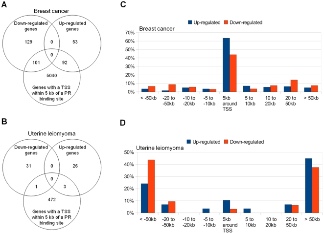
Principal findings: ChIP-sequencing revealed 31,457 and 7,034 PR-binding sites in breast cancer and uterine leiomyoma cells, respectively; 1,035 sites overlapped in both cell types. Based on the chromatin-PR interaction in both cell types, we statistically refined the consensus progesterone response element to G•ACA• • •TGT•C. We identified two striking differences between uterine leiomyoma and breast cancer cells. First, the cis-regulatory elements for HSF, TEF-1, and C/EBPα and β were statistically enriched at genomic RU486/PR-targets in uterine leiomyoma, whereas E2F, FOXO1, FOXA1, and FOXF sites were preferentially enriched in breast cancer cells. Second, 51.5% of RU486-regulated genes in breast cancer cells but only 6.6% of RU486-regulated genes in uterine leiomyoma cells contained a PR-binding site within 5 kb from their transcription start sites (TSSs), whereas 75.4% of RU486-regulated genes contained a PR-binding site farther than 50 kb from their TSSs in uterine leiomyoma cells. RU486 regulated only seven mRNAs in both cell types. Among these, adipophilin (PLIN2), a pro-differentiation gene, was induced via RU486 and PR via the same regulatory region in both cell types.

Conclusions: Our studies have identified molecular components in a RU486/PR-controlled gene network involved in the regulation of cell growth, cell migration, and extracellular matrix function. Tissue-specific and common patterns of genome-wide PR binding and gene regulation may determine the therapeutic effects of antiprogestins in uterine fibroids and breast cancer.


Conflict of interest statement

Competing Interests: The authors have declared that no competing interests exist.

Figures

Figure 1
Figure 1. Summary of PR-binding sites in uterine leiomyoma and breast cancer cells.
(A) Venn diagrams summarize the number of PR-binding sites in T47D breast cancer cells and uterine leiomyoma cells. (B) Validation of in vivo recruitment of PR to novel PR-binding regions identified by ChIP-seq. T47D and leiomyoma cells (from 5 different subjects) were treated with RU486 (10−6 M) or vehicle (ethanol) for 1 hour. ChIP was performed using anti-PR or nonspecific rabbit IgG, followed by real-time PCR. Data presented are expressed as fold enrichment of PR relative to IgG, and are the average of three to five independent ChIP experiments. (C) Correlation between the number of PR-binding sites in an individual chromosome (X-axis) to the chromosome length (Y-axis). (D) Correlation between the number of PR-binding sites (X-axis) with the number of TSSs (Y-axis) in an individual chromosome. (E) Distribution of PR-binding sites relative to their nearest TSSs. The percentages of all identified PR-binding sites in each cell type and region are shown.
Figure 2
Figure 2. Sequence logos of PRE consensus motifs.
The three known PRE motifs from TRANSFAC are shown in upper panels A (PR_01), B (PR_02), and C (PR_Q2). The consensus logo motifs derived using MEME analysis of ChIP-seq data from the 1,035 PR-binding sites common to both T47D breast cancer cells and leiomyoma cells are shown in lower panels of A, B, and C. Panel D shows the proposed consensus PRE motif based on the three TRANSFAC motifs and the discovered motifs from ChIP-seq data. For the consensus logos, the vertical axes (Bits) indicate the information content of the base frequency at that position. The horizontal axes refer to consensus site position.
Figure 3
Figure 3. Correlation between RU486-induced PR-binding and RU486-mediated gene expression.
Venn diagrams summarize the number of RU486-mediated genes with identified PR-binding sites within 5 kb from their TSSs in breast cancer cells (A) and leiomyoma cells (B). Distribution of PR-binding sites relative to TSSs of differentially expressed genes in T47D breast cancer cells (C) and uterine leiomyoma cells (D). T47D and leiomyoma cells were treated with vehicle (ethanol) or RU486 (10−6 M) for 6 hours and expression profiling was performed using Human HT-12 v4 Expression BeadChip arrays from Illumina. The percentage of all differentially regulated genes with PR-binding sites in each region is shown in the vertical axis. The distance relative to the TSS is shown in the horizontal axis.
Figure 4
Figure 4. Effect of RU486 treatment on the expression of PLIN2.
Expression of PLIN2 by real-time PCR in breast cancer T47D (A) and primary uterine leiomyoma cells (B). Serum-starved T47D or leiomyoma cells were stimulated with variable concentrations of RU486 (ranging from 10−9 to 10−5 M) or vehicle for 6 hours (C and D), or RU486 (10−6 M) or vehicle for 1, 3, 6, or 24 hours (E and F). PLIN2 mRNA levels were normalized to GAPDH expression. Results are reported as fold change compared with cells treated with vehicle only and represent the mean ± SEM of three independent experiments. Reference: star symbol, p<0.05 compared with vehicle treatment.

Similar articles

Cited by

References

    1. Shyamala G. Progesterone signaling and mammary gland morphogenesis. J Mammary Gland Biol Neoplasia. 1999;4:89–104. - PubMed
    1. Mulac-Jericevic B, Mullinax RA, DeMayo FJ, Lydon JP, Conneely OM. Subgroup of reproductive functions of progesterone mediated by progesterone receptor-B isoform. Science. 2000;289:1751–1754. - PubMed
    1. Conneely OM, Mulac-Jericevic B, Lydon JP. Progesterone-dependent regulation of female reproductive activity by two distinct progesterone receptor isoforms. Steroids. 2003;68:771–778. - PubMed
    1. Bulun SE, Cheng YH, Yin P, Imir G, Utsunomiya H, et al. Progesterone resistance in endometriosis: link to failure to metabolize estradiol. Mol Cell Endocrinol. 2006;248:94–103. - PubMed
    1. Kim JJ, Sefton EC. The role of progesterone signaling in the pathogenesis of uterine leiomyoma. Mol Cell Endocrinol. 2011 In press. - PMC - PubMed

Publication types

MeSH terms

Associated data